
Different Types of Microscopes: Almicro's Complete Guide to Modern Way
Table: Types of Microscope
- Compound Microscope
- Electron Microscope
- Simple Microscope
- Binocular Microscope
- Digital Microscope
- Dissecting Microscope
- Phase Contrast
- Medical and Research Microscope
Microscopes support daily laboratory work across medicine, education, industry, and research. Each microscope type serves a specific task. You select a microscope based on sample type, magnification needs, and workflow speed. This guide explains unique laboratory uses of major microscope types using clear and direct language. The focus stays on practical value and real laboratory application.
Compound Microscope
The compound microscope remains a core instrument inside laboratories. This microscope uses multiple lenses to reach higher magnification. Laboratories rely on this system for routine sample analysis.
Unique laboratory uses
• Blood smear examination during pathology testing
• Bacterial cell study during microbiology work
• Tissue section review during histology analysis
• Parasite identification in diagnostic labs
Why this microscope fits daily work
You gain consistent visual detail at magnifications from 40x to 1000x. Glass slides work easily with prepared stains. Medical labs depend on this microscope for daily reporting. Research labs use this microscope for cell counting and morphology review.
Example use
A pathology lab reviews red blood cell shape using a compound microscope during anemia screening. Clear lens alignment supports repeatable results across multiple samples.
Electron Microscope
The electron microscope operates at extreme magnification levels. Electron beams replace visible light. This structure supports nanoscale analysis.
Unique laboratory uses
• Virus structure study inside virology labs
• Organelle analysis during cell biology research
• Nanomaterial inspection inside material science labs
• Semiconductor surface review during industrial testing
Why laboratories invest in this system
This microscope reaches magnification above 100000x. Cellular membranes and viral capsids appear visible. Research institutions depend on this microscope for advanced structural data.
Example use
A pharmaceutical research lab studies viral protein structure using an electron microscope before vaccine formulation. Structural clarity supports drug design decisions.
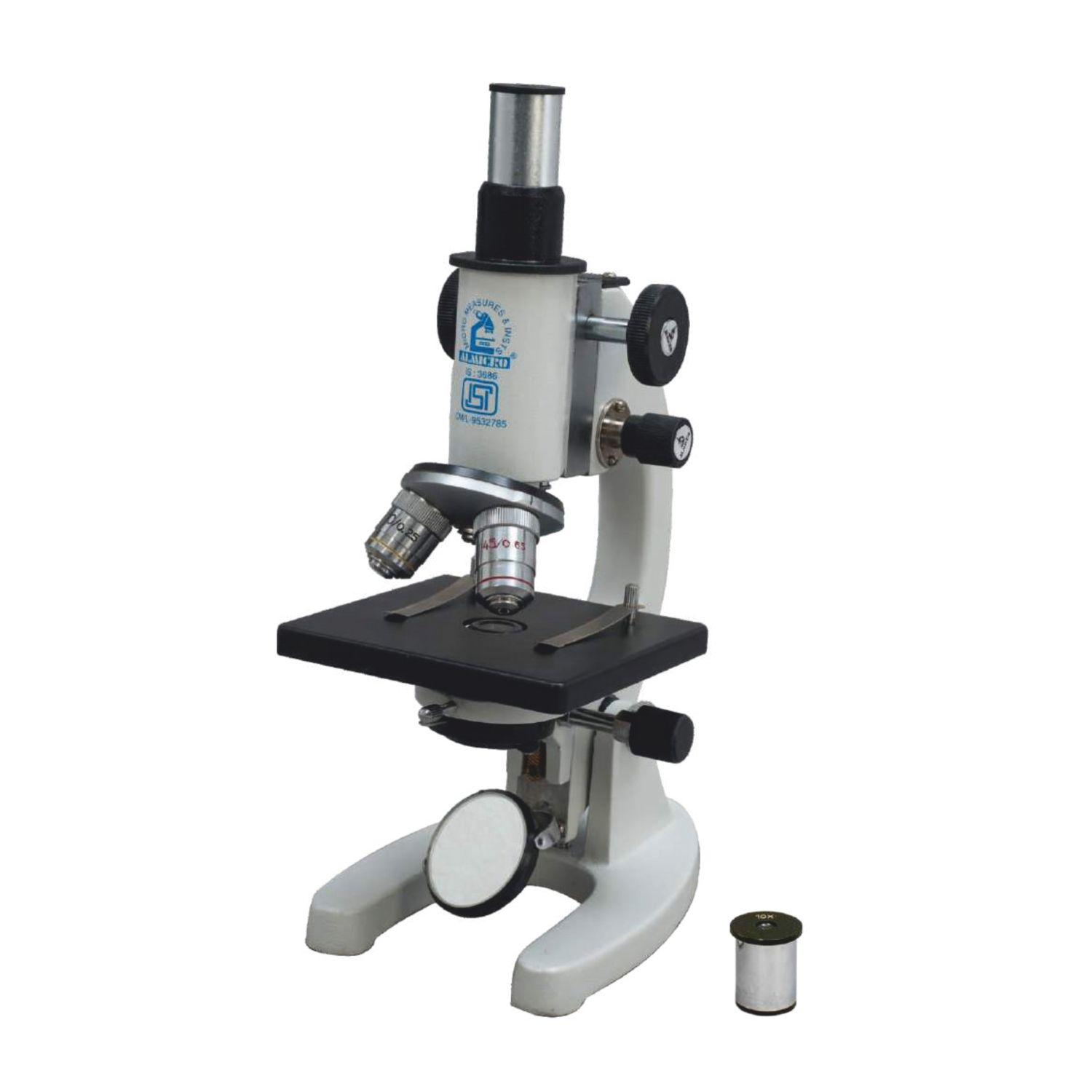
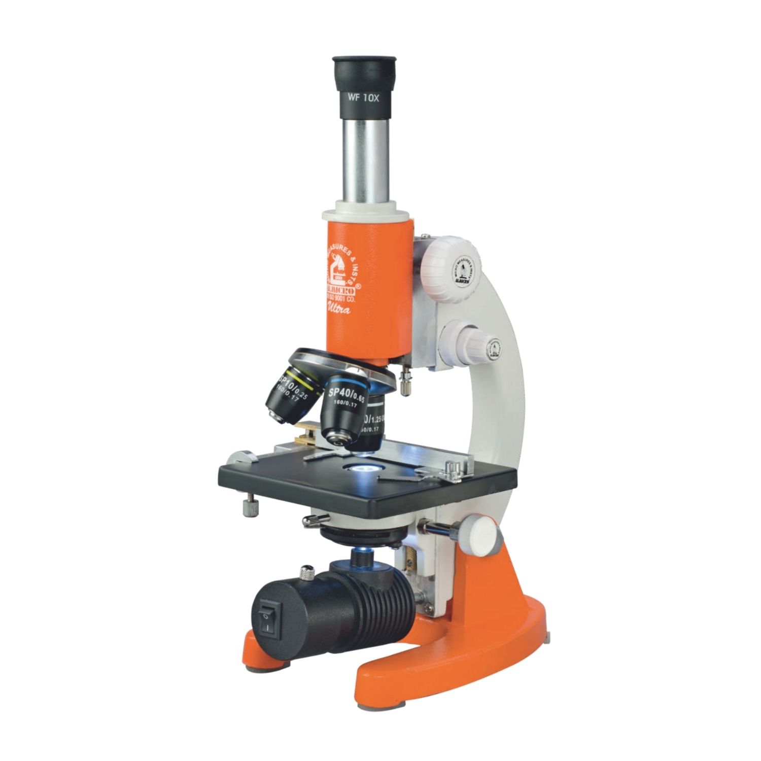
Simple Microscope

The simple microscope uses a single lens system. This microscope supports basic magnification tasks.
Unique laboratory uses
• Field sample review during environmental testing
• Crystal inspection during chemistry education
• Textile fiber checking during quality testing
• Quick specimen screening during training sessions
Why this microscope stays relevant
This microscope supports portability and speed. Students learn basic microscopy principles using this tool. Field technicians carry this microscope for rapid checks.
Example use
An environmental lab technician checks soil particles on site using a simple microscope during water contamination assessment.
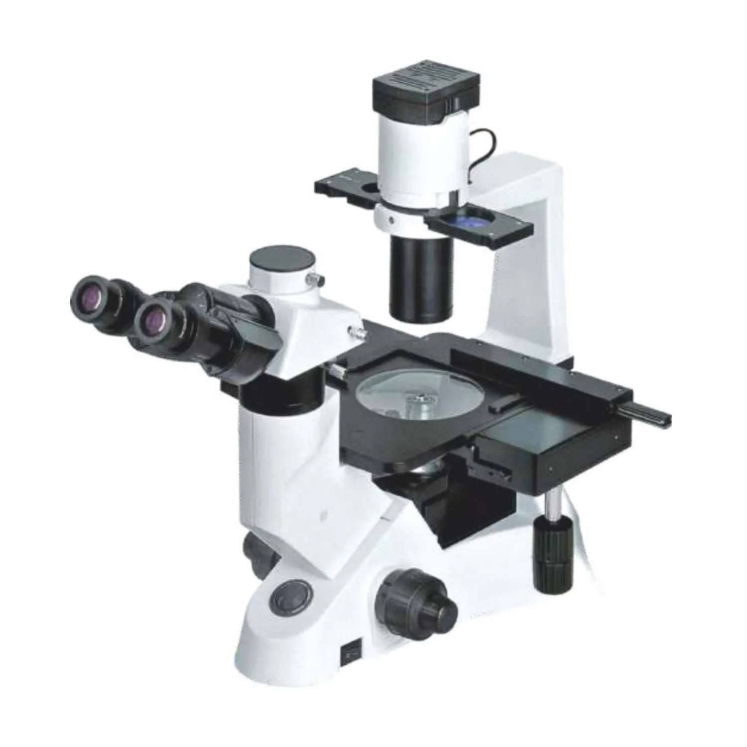
Binocular Microscope
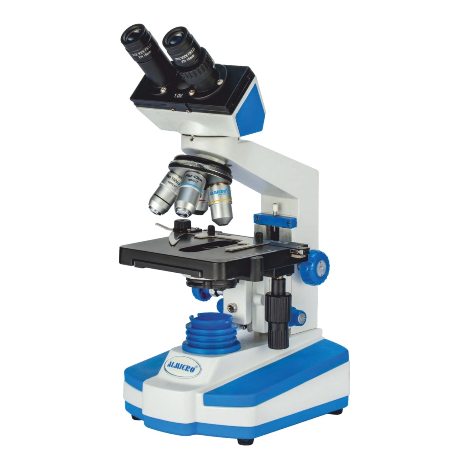
The binocular microscope uses two eyepieces. This design reduces eye strain during long sessions.
Unique laboratory uses
• Clinical slide analysis during long diagnostic shifts
• Academic teaching labs with multiple students
• Cytology labs reviewing large sample volumes
• Quality control labs handling repetitive inspection
Why this microscope improves workflow
Two eyepieces support natural viewing posture. Extended sessions become manageable. Laboratory staff stay focused without discomfort.
Example use
A diagnostic center screens hundreds of Pap smear slides daily using binocular microscopes to reduce operator fatigue.
Digital Microscope
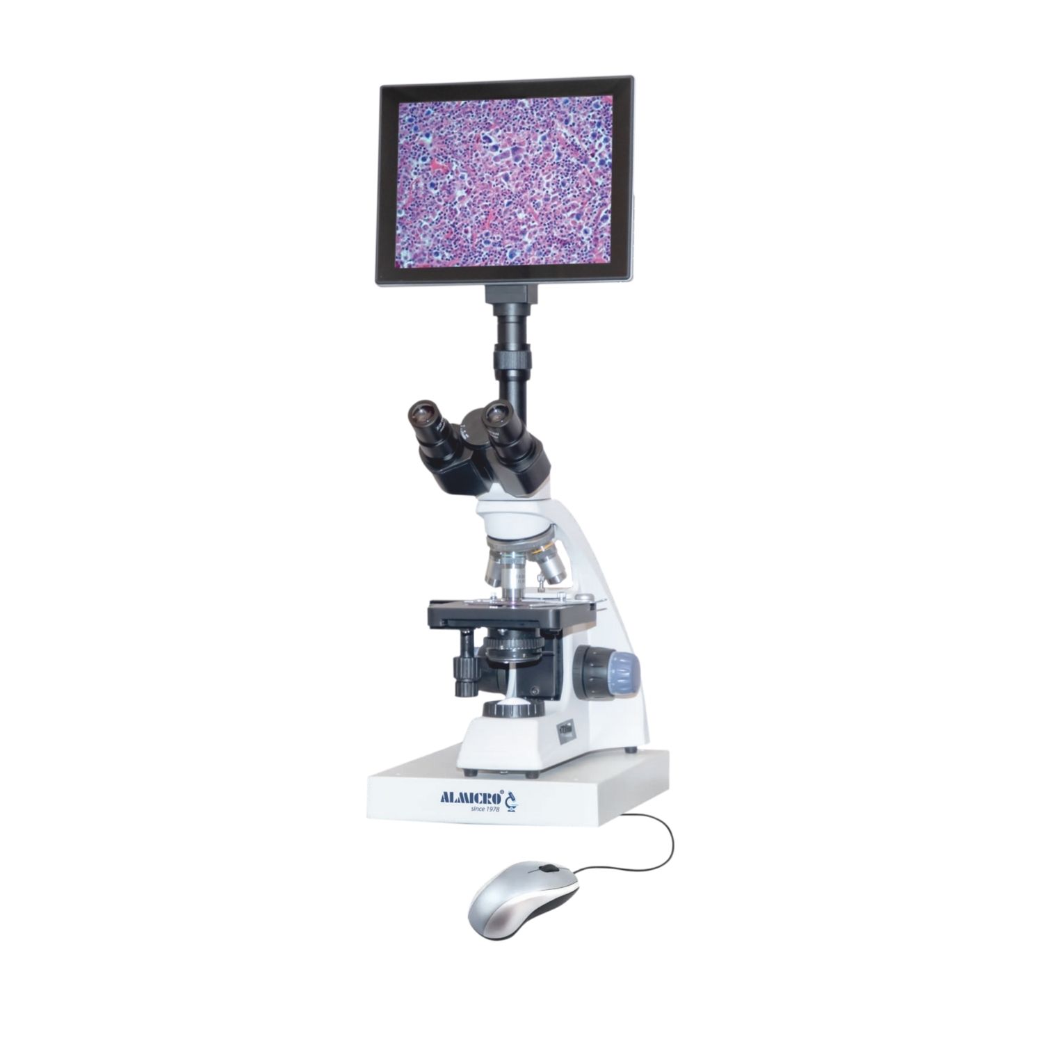
The digital microscope connects directly to screens. Images appear on monitors without eyepieces.
Unique laboratory uses
• Documentation during forensic examination
• Image capture for laboratory reports
• Remote teaching and training sessions
• Industrial inspection with image storage
Why this microscope fits modern labs
You record images instantly. Team members review samples together on screens. Data storage supports audit and training needs.
Example use
A forensic lab records fiber comparison images using a digital microscope during criminal investigation documentation.
Dissecting Microscope
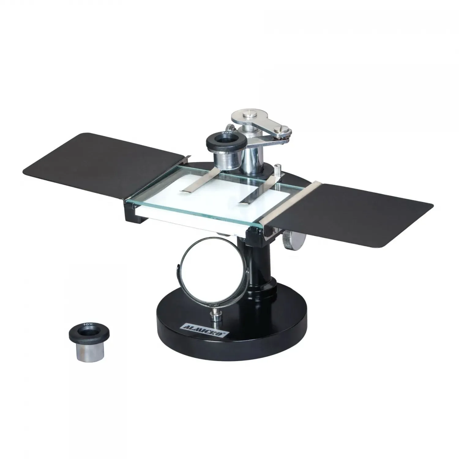
The dissecting microscope supports low magnification with a wide field view. Three dimensional viewing supports surface analysis.
Unique laboratory uses
• Insect dissection during entomology studies
• Circuit board inspection during electronics testing
• Plant anatomy review during botanical research
• Surgical training labs requiring depth perception
Why this microscope supports surface work
Depth view helps manual handling. Sample preparation stays minimal. Large specimens fit easily.
Example use
A biology lab dissects frog specimens using a dissecting microscope during anatomy coursework.
Phase Contrast Microscope
The phase contrast microscope supports transparent sample study without staining.
Unique laboratory uses
• Live cell culture monitoring
• Bacterial motility analysis
• Sperm analysis during fertility testing
• Tissue culture growth tracking
Why laboratories depend on this system
Cells remain alive during viewing. Structural contrast appears without chemical dyes. Research labs value this microscope during time sensitive experiments.
Example use
A cell biology lab monitors stem cell growth daily using a phase contrast microscope to avoid staining interference.
Medical Microscope
Medical microscopes serve diagnostic and surgical environments. These systems support patient focused workflows.
Unique laboratory uses
• Pathology slide review during disease diagnosis
• Hematology analysis for blood disorders
• Microbiology testing for infection detection
• Teaching hospitals for resident training
Why medical labs rely on this microscope
Clear optics support fast diagnosis. Ergonomic design suits extended clinical shifts. Hospitals integrate this microscope into daily patient care.
Example use
A hospital pathology lab identifies cancer cell margins using a medical microscope during biopsy evaluation.
Research Microscope

Research microscopes support advanced scientific investigation. Modular design allows configuration based on study needs.
Unique laboratory uses
• Fluorescence tagging during molecular biology
• Protein interaction analysis
• Advanced imaging for academic publications
• Multidisciplinary research environments
Why researchers select this system
Custom configurations support evolving projects. Imaging methods adapt to study design. Research funding often targets this equipment due to flexibility.
Example use
A university research lab studies neuron signaling using a research microscope fitted with fluorescence modules.
How You Choose the Right Microscope
Your selection process affects results quality and workflow speed. Focus on sample type and usage frequency.
Selection checklist
• Sample size and thickness
• Required magnification level
• Live sample handling needs
• Documentation requirements
• Operator training level
Laboratory impact
Correct selection reduces rework. Staff efficiency improves. Data consistency increases.
Laboratory Safety and Maintenance
Microscopes demand careful handling. Dust control and lens cleaning protect optical performance.
Best practices
• Cover microscopes after use
• Clean lenses with approved materials
• Store slides properly
• Schedule periodic calibration
Operational result
Maintenance routines support consistent output. Equipment lifespan extends. Laboratory downtime decreases.
Future Trends in Laboratory Microscopy
Laboratories continue adopting digital integration. Image sharing supports collaborative research. Automation reduces manual workload.
Emerging patterns
• Screen based analysis
• Image software integration
• Remote expert consultation
• Automated counting systems
Laboratory planning
Investment decisions shift toward flexible systems. Training focuses on digital interpretation skills.
Final Perspective
Microscopes support every laboratory discipline. Each type serves a defined purpose. You improve outcomes by matching microscope design to laboratory task. Knowledge of microscope uses guides smarter investment and better daily results.