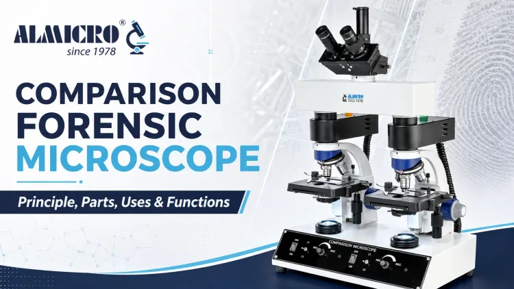
Comparison Forensic Microscope: Principle, Parts, Uses & Functions
A comparison forensic microscope is an optical instrument that lets an examiner view two specimens side by side in a single combined field to detect matching or differing microscopic features. Built around twin optical paths joined by a comparison bridge, it is a mainstay in forensic laboratories, where investigators need to place two pieces of evidence under identical magnification and lighting at the same time. Ballistics experts use it to match bullets and cartridge cases, while document examiners, hair and fiber analysts, and tool-mark specialists rely on the same side-by-side principle to build reliable, court-ready comparisons.
What Is a Comparison Forensic Microscope?
At its core, a comparison microscope is really two microscopes joined into one. Each specimen sits on its own stage, under its own objective lens, with its own illumination. Rather than forcing an examiner to look at one sample, note details, and then switch to the next, the instrument merges both images into a single eyepiece view, split down the middle or arranged side by side.
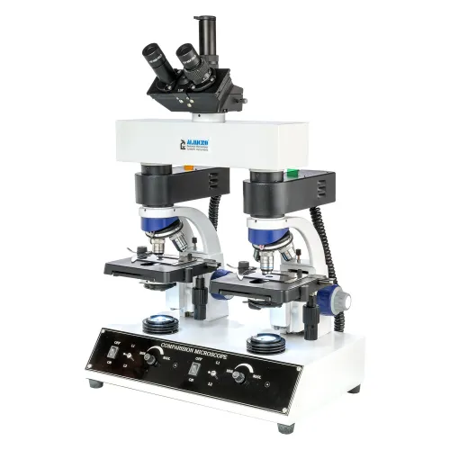
This split-field arrangement is what sets it apart from a standard compound microscope, which is built to examine only one specimen at a time. In forensic laboratories, that difference matters enormously. A firearms examiner comparing two bullets, or a trace-evidence analyst comparing two fibers, needs to see both samples under identical conditions at the same instant to draw a defensible conclusion.
Comparison Microscope Principle
The working principle behind a comparison microscope is straightforward, even though the optics inside it are precise. Two specimens are placed on separate stages, each with its own independent optical path: its own objective lens, focus control, and often its own illuminator.
Light from each specimen travels through its respective optical system and forms an image. The two images are then routed into a shared optical bridge, sometimes called a comparison bridge or comparison eyepiece assembly. This bridge combines the images optically, so the examiner sees a single combined field showing both specimens at once, typically separated by a thin dividing line down the center.
Specimen A + Specimen B → Separate optical systems → Comparison bridge → Combined split-field view → Examiner
Because both images appear under matching magnification and lighting, subtle similarities and differences in striations, edges, textures, and other microscopic features become easier to identify and document than they would be when switching between two separate microscopes.
Main Parts of a Comparison Microscope and Their Functions
A comparison microscope brings together several components that work in tandem to make simultaneous viewing possible.
| Part | Function |
|---|---|
| Dual stages | Hold the two specimens being compared |
| Objective lenses | Magnify each specimen independently |
| Illuminators | Provide dedicated lighting for each sample |
| Comparison bridge | Combines the two optical paths into one field |
| Eyepieces | Display the merged, split-field view |
| Focus controls | Allow each specimen to be focused separately |
| Stage controls | Position and align each specimen precisely |
| Camera port | Enables image capture for documentation |
| Magnification controls | Select the enlargement level needed for the comparison |
Together, these parts let an examiner move a bullet, fiber, or document fragment into exact alignment with its counterpart before drawing a conclusion.
Comparison Forensic Microscope Uses
Firearms and Ballistics
In forensic ballistics, the comparison microscope is a standard tool for comparing bullets, cartridge cases, firing-pin impressions, and breech-face marks left on ammunition components. Examiners align a recovered bullet against a test-fired sample to look for corresponding striation patterns.
Tool-Mark Examination
Cutting, scraping, and impact marks left by tools at a crime scene can be compared with marks produced by a suspected tool to assess whether they share corresponding microscopic features.
Hair and Fiber Comparison
The instrument is used to study the physical structure and characteristics of hair and fiber samples side by side, helping analysts note similarities and differences in color, texture, morphology, or damage.
Paint and Glass Examination
Paint chips and glass fragments can be compared for visible physical characteristics. Layer sequences, colors, surface features, and manufacturing differences may become clearer under matched magnification.
Questioned Documents
Where microscopy is appropriate, document examiners use comparison microscopy to study printing, ink deposition, paper fibers, and surface features side by side.
How Does a Comparison Microscope Help in Forensic Examination?
The real value of a comparison microscope lies in simultaneous observation. Because both specimens appear at the same magnification and within the same visual field, an examiner can compare microscopic features directly instead of relying on memory while switching back and forth between separate instruments.
This can make the comparison process more efficient and reduce the chance of overlooking a subtle difference. Many comparison microscope setups also support photographic or digital documentation, allowing the field used during examination to be captured and preserved as part of the case record for review or presentation.
Comparison Microscope Advantages and Disadvantages
| Advantages | Disadvantages |
|---|---|
| Allows simultaneous examination of two specimens | Requires trained and experienced operators |
| Supports direct side-by-side visual comparison | High-quality systems can be expensive |
| Makes fine surface details easier to compare | Accurate alignment and calibration are essential |
| Supports image capture and documentation | Interpretation still depends on expert judgment |
| Can save time compared with separate observation | Only suitable for evidence that can be compared microscopically |
Comparison Microscope vs Compound Microscope
It helps to see how a comparison microscope differs from the compound microscope found in most general laboratories.
| Feature | Comparison Microscope | Compound Microscope |
|---|---|---|
| Number of specimens | Two, viewed together | Usually one at a time |
| Main purpose | Direct side-by-side comparison | General magnification and observation |
| Optical paths | Dual | Single |
| Split-field view | Yes | No |
| Common use | Forensic science | Biology and general laboratory work |
| Evidence comparison | Purpose-built for direct comparison | Limited |
Both instruments have their place, but only the comparison microscope is purpose-built for matching two specimens against each other in a combined field.
Digital Comparison Forensic Microscope
Modern comparison microscopes increasingly integrate digital cameras, allowing examiners to capture still images and video of the combined field instead of relying on sketches or written notes alone. Side-by-side digital documentation makes it easier to archive a comparison as it appeared through the optical system, and many setups include measurement software for recording dimensions or annotating specific features.
This kind of digital record supports clearer case reports and makes it simpler to revisit a comparison if a finding is questioned. Manufacturers such as ALMICRO offer comparison microscope systems with digital camera compatibility and imaging options for laboratories that need dependable documentation alongside optical performance.
Comparison Forensic Microscope Notes: Key Points to Remember
- The instrument is designed for the simultaneous comparison of two specimens.
- Dual optical systems feed into a single combined field rather than two unrelated views.
- It is widely associated with forensic ballistics but also supports hair, fiber, tool-mark, paint, glass, and selected document examinations.
- Reliable results depend on proper calibration, suitable evidence, and trained interpretation.
- Digital systems can capture and preserve findings as part of the case record.
How to Choose a Comparison Forensic Microscope
Selecting the right comparison microscope for a laboratory comes down to several practical factors. Optical clarity and magnification range determine how much useful detail the instrument can resolve, while independent focusing and precise stage movement affect how easily two specimens can be aligned. Illumination control also matters because mismatched lighting between the optical paths can complicate a comparison.
- Optical quality: Check resolution, contrast, objective quality, and useful magnification range.
- Stage precision: Confirm that both specimens can be positioned and aligned accurately.
- Independent controls: Look for separate focusing and illumination adjustment for each optical path.
- Comparison bridge: Evaluate image alignment, field division, and viewing comfort.
- Digital compatibility: Check camera support, measurement tools, image capture, and reporting software.
- Ergonomics: Consider viewing posture and control placement for extended examinations.
- Service support: Confirm installation, calibration, training, spare parts, and after-sales assistance.
Laboratories weighing these criteria can evaluate manufacturers such as ALMICRO on optical quality, stage precision, illumination control, digital imaging compatibility, and after-sales support alongside other shortlisted comparison microscope systems.
Frequently Asked Questions
Choosing the Right Comparison Forensic Microscope for Laboratory Analysis
A comparison forensic microscope earns its place in a forensic laboratory through one core capability: putting two specimens under controlled, corresponding conditions and letting an examiner study them in a single combined field. That capability drives its role in ballistics, tool-mark analysis, and trace-evidence comparison. When evaluating a system, optical quality, stage precision, illumination control, and digital documentation options matter as much as brand reputation. ALMICRO's comparison microscope range is built with these forensic laboratory requirements in mind, offering the optical performance and support needed for dependable examination workflows.