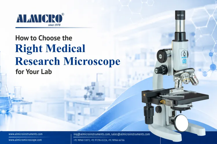
How to Choose the Right Medical Research Microscope for Your Lab?
Most procurement mistakes with MEDICAL RESEARCH MICROSCOPES don't happen at the ordering stage they happen months earlier, when someone signs off on a spec sheet without knowing what actually matters. Labs routinely overpay for optics they'll never use, get locked into non-compliant units that fail an audit, or get sold on a "1000x magnification" claim that hides a weak, unusable image. This guide gives you a procurement lens, not just an optical one the same precision-first thinking manufacturers like Almicro instruments apply when engineering research-grade optics from the ground up.
Below, we break down what separates a real research-grade system from a routine lab scope, map microscope types to actual sample workflows, and show you how to read a spec sheet so a sales rep can't upsell you on numbers that don't translate into usable data. New to the topic? Start with the fundamentals first:
Research-Grade vs Routine Microscope
What's the Difference?
A medical laboratory microscope used for routine diagnostics and a true research-grade system are built to solve different problems. Routine scopes are optimized for speed and consistency across repetitive slide checks. Research systems are optimized for image accuracy, reproducibility, and the ability to scale as your study design changes.
The difference shows up in three places on the spec sheet:
- Optics correction: Routine scopes typically use achromatic objectives, fine for basic viewing but prone to color fringing and edge distortion. Research-grade systems use plan-achromat or plan-apochromat objectives, correcting for both chromatic and field curvature errors across the entire field of view.
- Frame stability: Research imaging especially long-exposure fluorescence or time-lapse work demands a mechanically rigid frame and stage. Micro-vibrations that a routine scope tolerates will blur research-grade image capture.
- Upgrade paths: A routine microscope is usually a closed system. A research-grade frame is modularly built to accept fluorescence attachments, digital camera ports, or motorized stages later, without replacing the whole unit.
This is the distinction manufacturers like Almicro engineer around: building a base frame precise enough to support add-on modalities instead of forcing a full system replacement two years into a study.
Types of Medical Research Microscopes
Not every research question needs the same instrument. A biological research microscope built for live-cell imaging is a poor fit for fixed tissue histology, and vice versa matching type to sample is the first real procurement decision. Sourcing from a proven MEDICAL RESEARCH MICROSCOPES manufacturer in world markets matters here too, since it means your base system can genuinely expand into fluorescence or phase contrast modalities as your research scope grows, rather than hitting a hardware ceiling.
| Microscope Type | Best For | Sample Type | Contrast Method | Investment Tier |
|---|---|---|---|---|
| Compound Brightfield | General histology and cytology | Fixed, stained slides | Absorption / Staining | Entry |
| Phase Contrast / DIC | Live cell observation | Unstained live cells | Optical phase shift | Mid |
| Fluorescence | Protein and marker localization | Labeled cells and tissues | Excitation / Emission | Mid–High |
| Inverted | Live cell culture applications | Cells in flasks or culture dishes | Brightfield / Phase Contrast | Mid |
| Stereo | Dissection and sample preparation | Whole specimens | Reflected light | Entry–Mid |
Each type serves a distinct stage of research, from initial sample prep on a stereo scope to detailed marker-based imaging on a fluorescence system. The right starting point depends on your sample state live vs. fixed more than on budget alone.
Specification Checklist How to Read a Spec Sheet
Once you've matched microscope type to sample, the real filtering happens on the spec sheet and this is where most procurement teams get misled. A laboratory research microscope listing "1000x magnification" in bold but omitting numerical aperture is a red flag, not a selling point.
Resolution comes from Numerical Aperture (NA), not magnification.
Magnification just enlarges the image; NA determines whether fine detail is actually resolvable or just blown up and blurry. This single distinction separates usable research data from wasted budget. It's also why sourcing from established MEDICAL RESEARCH MICROSCOPES manufacturers in India matters for compliance-heavy labs regional manufacturers built for BIS and NABL-aligned procurement tend to document NA, objective class, and illumination specs transparently, rather than burying them behind headline magnification numbers.
| Parameter | Accept | Question |
|---|---|---|
| Numerical Aperture (NA) | Clearly listed per objective (e.g., 0.65, 1.25 Oil) | Only magnification is listed with no NA value. |
| Objective Class | Plan Achromat or Plan Apochromat clearly specified. | Only "Achromatic" is mentioned without correction type. |
| Illumination | Köhler illumination with adjustable LED and specified lux/CCT. | Only "Bright LED" is mentioned without lux or CCT details. |
| Camera Sensor | Resolution, sensor size, and frame rate are specified. | Described only as an "HD Camera" without sensor specifications. |
| Ergonomics | Adjustable eyepiece tilt and tension-adjustable focus are provided. | No information about comfort features for extended use. |
If a spec sheet can't answer the "Question" column clearly, treat it as incomplete not as a
Application-Specific Selection Matrix
Optical configuration should follow your workflow, not the other way around. A clinical research microscope chosen for histopathology will underperform badly in a live-cell IVF lab, and vice versa the table below maps common domains to their minimum viable specification.
| Application Domain | Recommended System Type | Min. Required NA | Primary Contrast Method | Digital Camera Required? |
|---|---|---|---|---|
| Histopathology & Biopsy | Compound Brightfield (Plan-Apochromat) | 0.65–0.85 | Brightfield / H&E Staining | Yes (Archiving & Telepathology) |
| Hematology & Cytology | Compound Brightfield with Oil Immersion | 1.25 (Oil) | Brightfield / Giemsa Staining | Recommended |
| Microbiology & Parasitology | Compound Brightfield / Darkfield | 0.65–1.25 | Brightfield / Darkfield | Optional |
| Live Cell Culture & IVF | Inverted Phase Contrast | 0.30–0.55 | Phase Contrast | Yes (Time-Lapse Imaging) |
| Pharma Quality Control | Stereo / Compound Brightfield | 0.10–0.65 | Brightfield / Polarized Light | Yes (Documentation & Audit Trail) |
Notice the NA floor rises sharply for oil-immersion hematology work and drops for low-power QC inspection. Matching this correctly upfront avoids both underpowered optics and unnecessary spend on capability you won't use.
Compliance & Accreditation Requirements
A technically strong medical microscope for pathology is worthless in an accredited lab if it can't pass documentation review. Compliance isn't a formality, it's a procurement filter that should run parallel to your optical spec comparison, not after it.
Four frameworks typically govern lab microscope procurement:
- ISO 13485 — Confirms the manufacturer's quality management system for medical device production, not just the product itself.
- IVD / EU IVDR / FDA — Relevant when the microscope is part of a diagnostic workflow feeding clinical decisions, not pure research use.
- NABL / CAP / ISO 15189 — Governs lab accreditation itself; auditors will ask for traceable calibration records tied to the specific instrument, not a generic certificate.
Auditors don't just check the microscope, they check its calibration paper trail.
This is where sourcing decisions matter operationally. Established medical research microscopes manufacturers in India Almicro among them typically provide traceable stage micrometer calibration certificates and regional service support, which shortens the gap between an audit finding and a resolved corrective action.
Investment Tiers Budget Planning Without Guesswork
Budget planning breaks down when every vendor quote bundles different optics, illumination, and software into one number. Separating systems into tiers makes the comparison honest.
| Tier Level | Optics Class | Illumination Type | Imaging & Software Suite | Best Suited For |
|---|---|---|---|---|
| Tier 1 — Standard | Achromat / Plan-Achromat | LED (Fixed Intensity) | Basic Image Capture Software | Routine Diagnostics and Teaching Laboratories |
| Tier 2 — Mid-Range | Plan-Achromat / Semi-Apochromat | Adjustable LED with Köhler Illumination | Measurement and Basic Analysis Software | Clinical Research, Hematology, and Quality Control |
| Tier 3 — Advanced Core | Plan-Apochromat | High-CRI LED, Fluorescence-Ready | Advanced Analysis Suite with Multi-User Licensing | Core Imaging Facilities and Publication-Grade Research |
Tier 3 systems earn their premium through correction quality and expandability, not brand markup. A Tier 2 system with a genuine upgrade path is often the better long-term buy than a Tier 3 system you won't fully use for two years.
Hidden Costs What Isn't in the Initial Quote
The purchase price of a medical imaging microscope is rarely the real cost. Total Cost of Ownership (TCO) shows up over the following 3–5 years, and most quotes are silent on it.
Before signing, confirm these five line items are addressed — not assumed:
- AMC/CMC — Annual or Comprehensive Maintenance Contract terms and included visit count
- Calibration & Validation — Frequency, certificate format, and whether it's traceable to a national standard
- Software Licensing — Per-seat vs. per-device licensing, and renewal cost after year one
- Optical Consumables — Immersion oil, cleaning kits, replacement bulbs/LED modules, filter cubes
- Installation & Training — On-site setup, operator training hours, and whether refresher training is chargeable
A quote missing three or more of these items isn't a lower price — it's an incomplete one.
Frequently Asked Questions (FAQs)
Q1: Is higher magnification always better for research use?
No. Magnification without adequate Numerical Aperture just enlarges a blurry image. NA determines whether structural detail is actually resolvable.
Q2: Can a standard compound microscope be upgraded to fluorescence later?
Only if the base frame was designed for it. Confirm filter cube compatibility and light path access before purchase, not after the research need arises.
Q3: Why are plan-apochromat objectives priced significantly higher than plan-achromats?
They correct chromatic aberration across three or more wavelengths instead of two, and correct field curvature more precisely — critical for publication-grade imaging and multi-color fluorescence work.
Q4: LED or halogen illumination — does it matter for research imaging?
LEDs offer stable color temperature, longer lifespan, and lower heat output, all of which matter for live-cell and long-exposure imaging. Halogen is largely legacy at this point.
Q5: Does buying from a local medical research microscopes manufacturer actually matter?
Yes for calibration turnaround, spare parts availability, and compliance documentation aligned to regional accreditation bodies like NABL. It reduces downtime risk more than most buyers expect.
Industry Engagement & Certification Standards
Beyond specs and compliance frameworks, it's worth checking how visible and certified a manufacturer actually is. Almicro Instruments maintains an active exhibition presence across India, Asia, the Middle East, Africa, and Europe, including a recent showcase at an exhibition in Gandhinagar, Gujarat, where its team engaged directly with labs, research centers, and educational institutions.
On the certification side, Almicro's published records include ISO 9001, ISO 13485, CE marking, and US FDA registration documentation alongside NSF/ANSI 49 and EN 12469 for its biosafety cabinet range. These don't replace your lab's own accreditation (NABL, CAP, ISO 15189), but they do confirm the manufacturer's own quality processes have been externally reviewed.
Conclusion & Strategic Procurement Roadmap
Choosing the right medical research microscope setup comes down to three filters: verify Numerical Aperture before magnification, confirm compliance documentation before signing, and calculate TCO before comparing sticker prices. Skip any one of these, and the "savings" on the quote resurface later as downtime, audit findings, or a system you've outgrown in a year.