
What is Microscope ?
Microscopes are powerful instruments that enable us to see the minute world, revealing intricate features that are not visible to the unaided eye. They are indispensable in biology, medicine, materials science, and forensics. This blog post will take you through the amazing world of microscopes, discussing their meaning, operating principles, types, main characteristics, and advanced capabilities. Eventually, you will get a thorough knowledge of these mighty instruments for enlarging and investigating the unseen world.
Types of Microscope
1. Simple Microscope

Definition
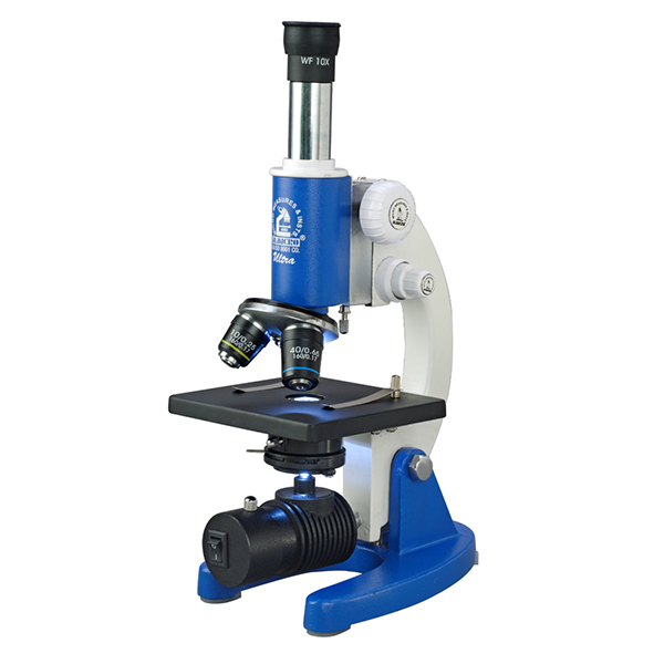
A Simple Microscope uses a single convex lens to magnify small objects. It produces an enlarged, upright, virtual image, similar to a magnifying glass. Though it offers lower magnification (10x–20x), it provides a direct and clear view, making it ideal for basic observation or dissection work.
Diagram Concept:

Label the convex lens as the single magnifying element.
Working Principle (Pointwise):
-
Place the specimen on a flat surface or stage.
-
Illuminate it with natural or artificial light.
-
Position the object within the focal length of the convex lens.
-
The convex lens bends the light rays.
-
The viewer sees an enlarged, virtual image of the specimen.
2. Compound Microscope
Definition
A compound microscope is an optical instrument that uses two lens systems—objective lenses and an eyepiece—to magnify small specimens. It works with visible light and produces a two-dimensional, inverted, and magnified image. Compound microscopes are widely used in biology, pathology, microbiology, and educational laboratories for observing thin and transparent samples such as cells and tissues.
Diagram Concept

Labels:
Eyepiece (Ocular Lens), Objective Lenses, Fine Focusing Knob, Stage, Condenser, Light Source (Mirror/Lamp), Coarse & Fine Adjustment Knobs<
Concept Explanation:
The objective lens creates a magnified real image of the specimen. The eyepiece further magnifies this image to form the final virtual image visible to the observer.
Working Principle (Pointwise)
-
A built-in light source or mirror directs visible light toward the specimen.
-
The condenser lens concentrates light onto the specimen placed on the stage.
-
Light passes through the thin, transparent specimen.
-
The objective lens produces an inverted, magnified, real image.
-
The eyepiece lens magnifies this real image again.
-
The observer sees a highly enlarged virtual image.
-
Total magnification = Objective magnification × Eyepiece magnification
3. Binocular Microscope

Definition:
A binocular microscope is a type of optical microscope that has two eyepieces, allowing you to view the specimen with both eyes instead of one. This makes observation more comfortable, reduces eye strain, and gives a better 3D-like visual experience.
Binocular Microscope Diagram

Detailed Working Principle
Light Illuminates the Specimen
The light source or mirror sends light through the specimen on the slide.
Objective Lens Forms a Real, Magnified Image
The objective lens collects light from the specimen and produces a real, enlarged, inverted image inside the microscope tube.
Eyepieces Magnify Again
Each eyepiece lens magnifies this real image further, forming a virtual, larger image that your eyes
Both Eyes View the Same Image
Even though there are two eyepieces, they both receive the image from the same objective lens system, giving:
-
Comfortable viewing
-
Better depth effect
-
Reduced eye strain
4. Medical Microscope

Definition
A medical microscope is a professional-grade compound or fluorescence microscope designed for diagnostic use. It is optimized for examining blood smears, tissues, and microorganisms. With ergonomic design and oil immersion objectives, it supports long laboratory sessions and accurate visual analysis in hospitals and research centers.
Diagram Concept:

Labels – Binocular Eyepieces, High-Quality Objective (100x Oil Immersion), Stage.
Working Principle (Pointwise):
- Prepare and stain biological specimens for better visibility.
- Use transmitted light to illuminate thin sections.
- Apply oil immersion for high-power objectives to increase resolution.
- Use phase contrast or darkfield for live, unstained samples.
- Observe and analyze for diagnostic results.
5. Digital Microscope
Definition
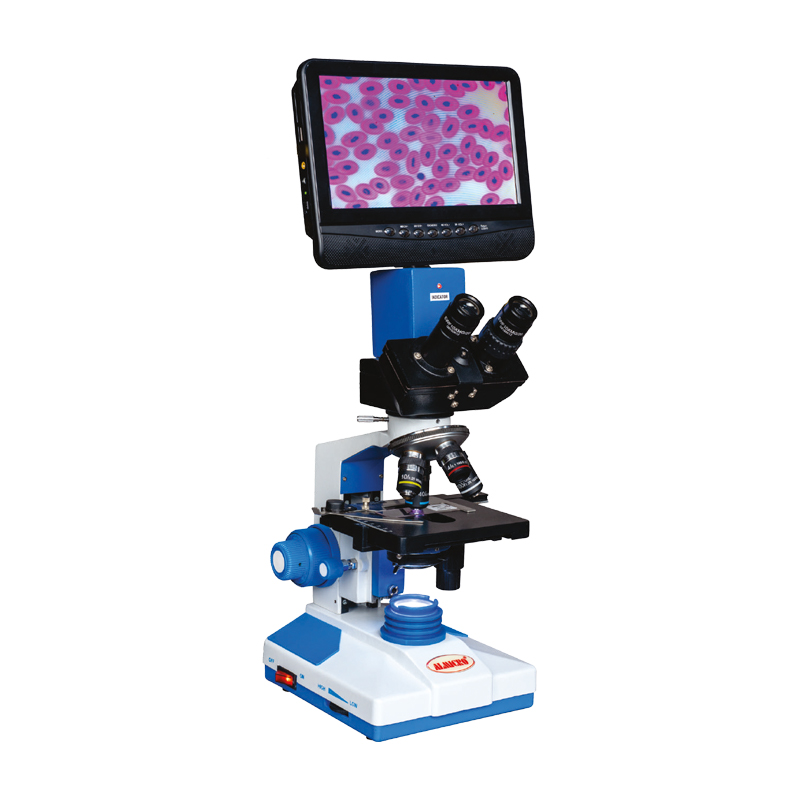
A digital microscope combines optical lenses with a digital camera (CCD or CMOS sensor). It captures magnified images and displays them on a computer or built-in screen, eliminating the need for eyepieces. It is used in research, education, and industry for documentation, analysis, and comfortable viewing.
Diagram Concept:

Labels – Digital Camera Sensor, Display Monitor, Stage, Objective Lens.
Working Principle (Pointwise):
- Illuminate the specimen and focus using the objective lens.
- The real image is projected onto a digital sensor.
- The sensor converts light into electronic signals.
- The processor transforms these signals into pixels.
- The digital image is displayed on a monitor.
- Software allows image capture, measurement, and sharing.
6. Stereo Microscope (Dissecting Microscope)
Definition
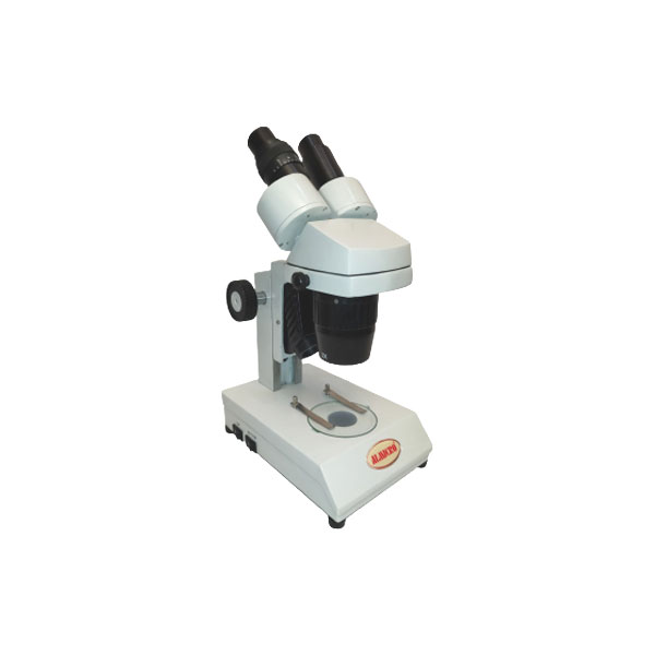
A Stereo Microscope provides a three-dimensional view of large, opaque specimens using two objective lenses and two eyepieces. Each eye receives a slightly different image, creating depth perception. It is used in dissection, electronics, and quality control, with magnification between 10x and 40x.
Diagram Concept:

Labels – Dual Objective Paths, 3D View, Reflected Light Illumination.
Working Principle (Pointwise):
- Illuminate the specimen surface using reflected light.
- Two separate optical paths capture images from different angles.
- Light from both paths reaches the eyepieces separately.
- The brain merges both views, giving a 3D image.
- The large working distance allows manipulation under magnification.
7. Light Microscope (General Type)

Definition
A Light Microscope uses visible light and glass lenses to magnify transparent specimens. It includes both simple and compound models. While its resolution is limited by the wavelength of light, it remains one of the most used and accessible tools for biological and educational studies.
Diagram Concept:

Labels – Visible Light Source, Glass Lenses, Stage, Eyepiece.
Working Principle (Pointwise):
- The specimen is illuminated with visible light.
- The light passes through or reflects off the specimen.
- Convex lenses bend (refract) light to enlarge the image.
- Objective and eyepiece lenses work together for magnification.
- Image contrast depends on light absorption and scattering.
8. Fluorescent Microscope

Definition
A Fluorescent Microscope uses high-intensity light (often ultraviolet or blue) to excite fluorescent dyes in a sample. These dyes emit light at longer wavelengths, allowing visualization of specific cellular structures with high contrast. It is crucial for studying proteins, cells, and biological interactions.
Diagram Concept:
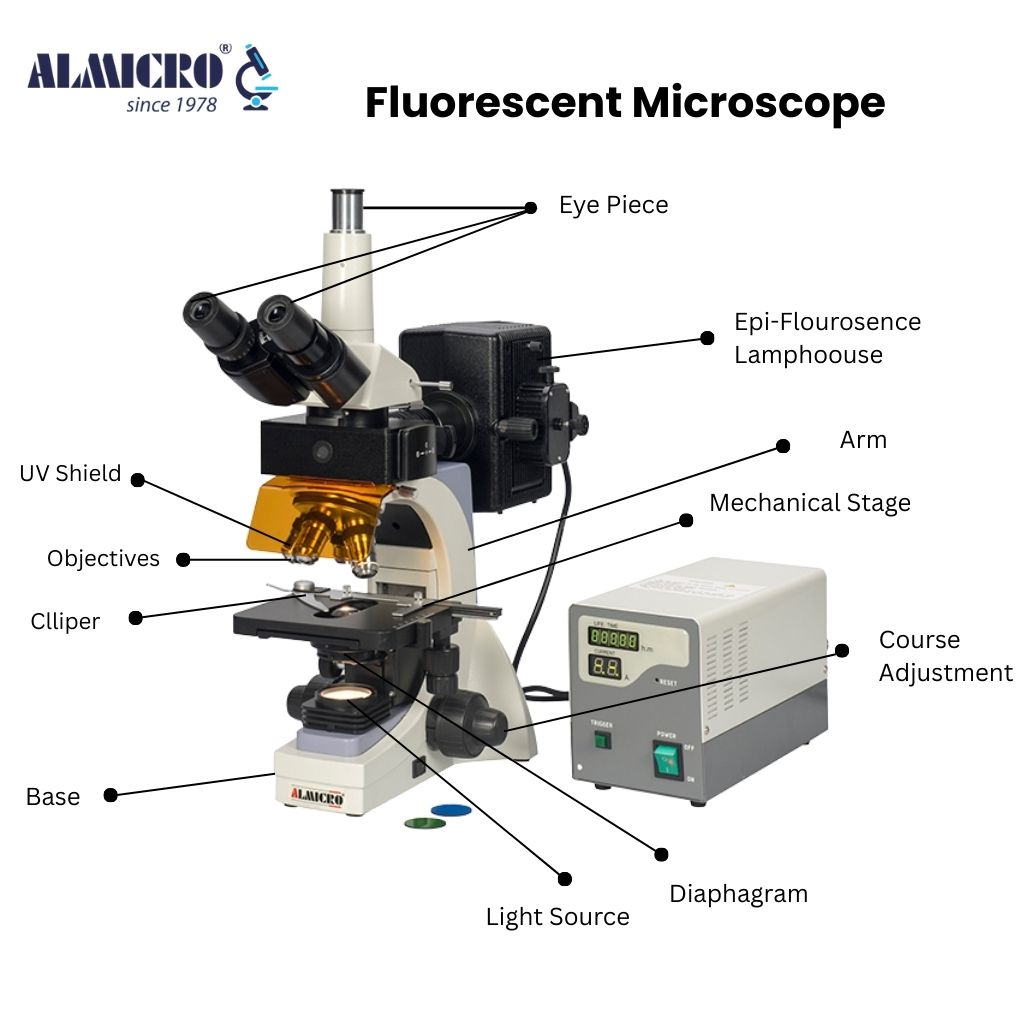
Labels – Excitation Light Source, Dichroic Mirror, Emission Filter, Detector.
Working Principle (Pointwise):
- Excitation light of short wavelength strikes the specimen.
- The specimen is stained with fluorescent dyes (fluorophores).
- Fluorophores absorb and emit light at different wavelengths.
- A dichroic mirror separates emitted and excitation light.
- Emitted light forms a bright fluorescent image against a dark field.
9. Confocal Microscope (Laser Scanning Microscope)

Definition
A Confocal Microscope scans specimens point-by-point using a focused laser beam. A pinhole filter blocks out-of-focus light, creating sharp optical sections. This allows precise 3D imaging of thick fluorescent samples, widely used in cell biology, materials science, and nanotechnology.
Diagram Concept:

Labels – Laser Source, Scanning Mirrors, Pinhole, Detector.
Working Principle (Pointwise):
- A laser beam is focused on a single point on the specimen.
- Scanning mirrors move the beam across the sample.
- Fluorescence from the focused point is collected.
- Out-of-focus light is blocked by a pinhole.
- The detector records the in-focus signal.
- Computer software reconstructs 2D or 3D images from scans.
10. Digital Light Microscope (Hybrid Type)

Definition
A Digital Light Microscope merges traditional optical viewing with digital capture. A digital camera attached to the trinocular head records images while the user simultaneously observes through eyepieces. It is used for research, documentation, and quality inspection in scientific and industrial applications.
Diagram Concept:

Labels – Trinocular Port, Digital Camera, Eyepiece, Computer Display.
Working Principle (Pointwise):
- Light passes through the specimen and lens system as in a compound microscope.
- A prism diverts part of the light to the camera.
- The camera captures digital images.
- Images are displayed on a computer in real time.
- Data is stored or analyzed through imaging software.
Frequently Asked Questions (FAQs) – Microscope
1. What is a microscope used for?
A microscope is used to observe very small objects such as cells, tissues, microorganisms, and material structures that cannot be seen with the naked eye. It is widely used in biology, medicine, research, education, and industrial analysis.
2. What is the basic working principle of a microscope?
A microscope works on the principle of magnification using lenses. Light passes through or reflects from the specimen, and convex lenses refract the light to produce an enlarged image that the human eye can clearly observe.
3. What is the difference between a simple and compound microscope?
A simple microscope uses a single convex lens and provides low magnification, similar to a magnifying glass. A compound microscope uses two lens systems—objective and eyepiece—to achieve much higher magnification and better resolution for observing cells and microorganisms.
4. Why is a binocular microscope better for long use?
A binocular microscope allows viewing with both eyes, which reduces eye strain and provides more comfortable and natural observation. It also offers a better depth effect, making it ideal for extended laboratory work.
5. What type of microscope is used in medical laboratories?
Medical laboratories commonly use compound and medical-grade microscopes with oil immersion objectives. These microscopes are designed for examining blood smears, tissues, bacteria, and other diagnostic samples with high accuracy.
6. What is a digital microscope and how does it work?
A digital microscope uses a camera sensor instead of eyepieces to capture magnified images. The image is displayed on a computer or built-in screen, allowing easy viewing, image storage, measurement, and sharing.
7. What is a stereo microscope mainly used for?
A stereo microscope is used for observing larger, opaque objects in three dimensions. It is commonly used in dissection, electronics inspection, entomology, and quality control because it provides depth perception and a large working distance.
8. What is the main advantage of a fluorescent microscope?
A fluorescent microscope allows visualization of specific cellular components using fluorescent dyes. It provides high contrast images and is especially useful in cell biology, molecular biology, and medical research.
9. Where can I buy a microscope?
You can buy high-quality microscopes directly from Almicro (Micro Measure), a trusted microscope manufacturer in India since 1978.
We offer all types of microscopes, including biological, stereo, digital, and laboratory microscopes for education, research, and medical use.
10. What is a digital light microscope (hybrid type)?
A digital light microscope combines traditional optical viewing with digital image capture. Users can observe samples through eyepieces while simultaneously recording images using a digital camera for analysis and documentation.
11. Which microscope is best for students and education?
For students, a compound light microscope is the best choice. It is easy to use, affordable, and suitable for studying basic biology concepts such as cells, tissues, and microorganisms.
12. What factors should be considered when choosing a microscope?
Key factors include magnification range, resolution, type of specimen, application (education, medical, research), viewing comfort (monocular or binocular), and whether digital imaging is required.
Related Article
2. What is a Simple Microscope