Trinocular Microscope: Working Principle, Diagram, Uses and Camera Setup Explained
A binocular microscope lets only one person examine a specimen at a time. That limits teaching, documentation and collaboration in modern laboratories. A trinocular microscope solves this by adding a third optical port that connects a camera. The view seen through the eyepieces can then be captured, recorded and shared. This article covers the working principle, parts diagram, laboratory uses and camera setup. Almicro Instruments, based in Ambala Cantt, India, supplies microscopes to medical colleges, pathology laboratories and research institutes. It is known for consistent optical quality and a broad product range.
What Is a Trinocular Microscope?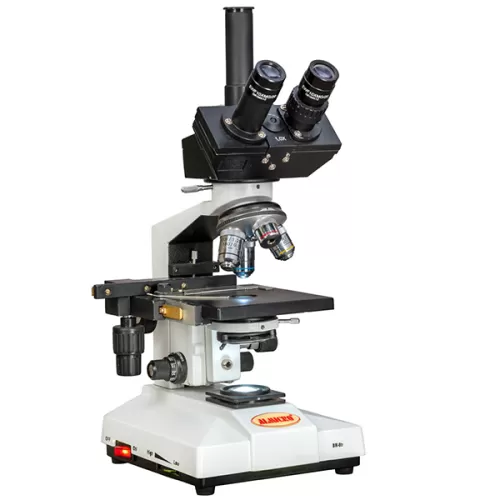
A trinocular microscope is a compound microscope with two eyepieces for direct viewing and a third, vertical port for mounting a camera. An observer can view a specimen while images or video are captured digitally at the same time.
Trinocular models belong to the compound microscope family, which uses multiple lenses to magnify small specimens. A monocular microscope has a single eyepiece and suits basic observation. A binocular microscope has two eyepieces, which reduces eye strain during long sessions. A trinocular microscope keeps the binocular viewing head and adds a photo tube above it, making it the standard choice for digital imaging.
Trinocular Microscope Working Principle
The trinocular microscope working principle combines standard compound optics with a beam splitter. The splitter shares the image between the observer and the camera.
How Light Travels Through the Microscope
Light from the illumination source passes through the condenser, which focuses it onto the specimen. The objective lens collects the light transmitted through the specimen and forms a magnified primary image. That image travels through the optical head to the prism and then to the eyepieces.
Total magnification is the objective magnification multiplied by the eyepiece magnification. A 10x eyepiece with a 40x objective gives 400x.
Role of the Beam Splitter in the Trinocular Head
Inside the trinocular head, a prism divides the optical path between the eyepieces and the camera port. Many models offer switchable light-split settings:
- 100:0 – all light goes to the eyepieces
- 50:50 – light is shared equally
- 0:100 – all light goes to the camera, useful for low-light imaging
Parfocal alignment keeps a specimen in focus on both the eyepieces and the camera sensor, so the user does not need to refocus. Image resolution depends mainly on the numerical aperture of the objective lens.
Trinocular Microscope Diagram and Parts Explained
A trinocular microscope diagram shows how the viewing head, objectives, stage and illumination system fit together. The third port on top of the head is what distinguishes it from a binocular design.
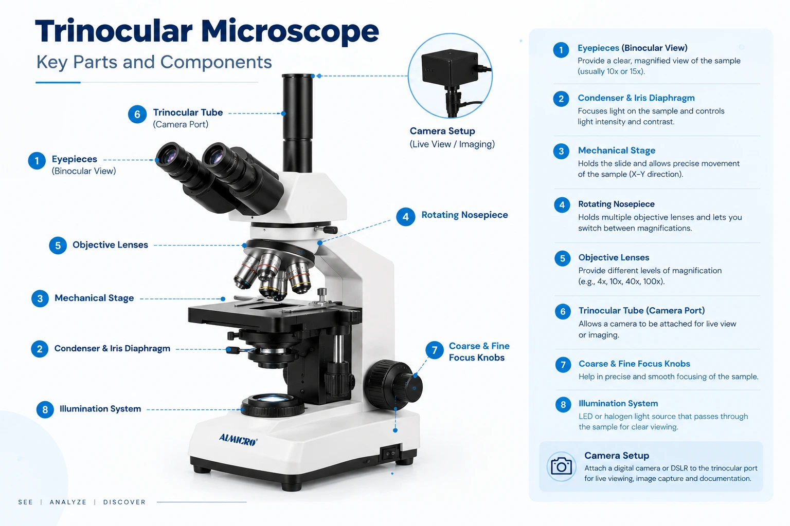
Alt text: Trinocular microscope diagram showing eyepieces, trinocular port, objectives and stage
| Part | Function |
|---|---|
| Eyepieces (10x) | Magnify the image for viewing |
| Trinocular port | Mounts the camera via C-mount |
| Beam splitter prism | Divides light between eyepieces and camera |
| Revolving nosepiece | Holds and rotates objective lenses |
| Objective lenses (4x–100x) | Provide primary magnification |
| Mechanical stage | Holds and moves the slide |
| Abbe condenser | Focuses light on the specimen |
| Coarse/fine focus knobs | Sharpen the image |
| LED illumination | Provides even, adjustable light |
Trinocular Microscope Uses in Labs and Industries
The main trinocular microscope uses involve observing and documenting specimens at once:
- Pathology and histopathology: slide documentation and second opinions
- Medical and dental colleges: live classroom display on screens
- Research and biotech laboratories: image capture for publications and time-lapse studies
- Pharmaceutical and industrial quality control: inspection records for audits
- Microbiology, food and environmental testing: sample analysis backed by visual evidence
- Telepathology: remote diagnosis and consultation
Because the image can be stored, compared and shared, the instrument supports both academic work and regulated testing.
Setting Up a Trinocular Microscope with Camera
A trinocular microscope with a camera needs three things: a suitable camera, a matching adapter and imaging software.
Choosing the Right Microscope Camera
Cameras use either CMOS or CCD sensors. Resolution typically ranges from 5MP to 20MP, and frame rate determines how smooth live video appears. Output options include USB, HDMI and Wi-Fi. HDMI suits classroom projection, while USB suits direct computer capture.
C-Mount Adapter and Reduction Lens
The camera attaches to the trinocular port through a C-mount adapter. A 0.5x reduction lens is commonly used so the camera's field of view matches what the eyepieces show. Without it, the sensor captures only a small central part of the image.
Imaging Software and Documentation
Imaging software provides live display, measurement tools, image annotation and report archiving. These features turn a microscope into a complete digital documentation system.
Trinocular vs Binocular Microscope: Which One Should You Choose?
| Feature | Binocular | Trinocular |
|---|---|---|
| Viewing ports | 2 | 2 + camera port |
| Camera support | Limited / eyepiece adapter | Dedicated port |
| Cost | Lower | Moderate |
| Best for | Routine viewing | Imaging, teaching, documentation |
Verdict: choose a trinocular microscope if your lab needs to record, teach or share images.
How to Choose a Trinocular Microscope Manufacturer in India
Buyers evaluating a trinocular microscope manufacturer in India should check:
- Plan achromatic objectives and overall optical clarity
- Camera and adapter compatibility
- Warranty, calibration and after-sales service
- Capacity for bulk or institutional supply
Almicro Instruments (Micro Measures & Instruments), based in Ambala Cantt, meets these criteria with more than 350 microscope models. Its range covers compound, binocular, trinocular, stereo, digital, metallurgical, polarizing, fluorescence and inverted types. It serves customers across India and in export markets, and it is a trusted source for institutions that need consistent quality at scale. Explore the trinocular microscope range
Frequently Asked Questions
What is a trinocular microscope used for?
It is used for viewing specimens while simultaneously capturing images or video through a camera for documentation, teaching and diagnosis.
Can any camera be attached to a trinocular microscope?
No. The camera needs a compatible C-mount adapter and suitable sensor size; dedicated microscope cameras give the best results.
What is the difference between binocular and trinocular microscopes?
A binocular microscope has two eyepieces only; a trinocular microscope adds a third port for camera mounting, enabling image capture and projection.
What magnification does a trinocular microscope have?
Typically 40x to 1000x, calculated by multiplying a 10x eyepiece by objectives from 4x to 100x.
Why a Trinocular Microscope Is Now a Lab Essential
A trinocular microscope combines direct viewing with image capture, recording and sharing in one instrument. That is why medical colleges, pathology laboratories and research institutes now treat it as standard equipment. Almicro Instruments is a trusted trinocular microscope manufacturer in India, with a wide range built for teaching, diagnosis and documentation. Request a quotation or download the Almicro microscope catalogue today.