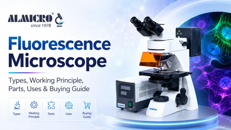
Fluorescence Microscope: Types, Working Principle, Parts, Uses & Buying Guide
A fluorescence microscope uses fluorescent dyes, proteins, or naturally fluorescent molecules to reveal selected structures within a specimen. It illuminates the sample with an excitation wavelength and detects the longer-wavelength light emitted by fluorophores. This produces high-contrast images of specific cells, proteins, microorganisms, or molecular targets. This guide explains the fluorescence microscope principle, light path, main parts, working steps, uses, applications, advantages, limitations, and buying considerations.
What Is a Fluorescence Microscope?
A fluorescence microscope is an optical instrument used to observe samples containing fluorophores. These fluorescent molecules absorb light within a specific excitation range and emit light at a longer wavelength. The microscope separates the emitted fluorescence from the stronger excitation light to form the final image.
 Read on Medium Complete Fluorescence Microscopy Guide ?
Important distinction: A fluorescence light microscope is another name for an optical fluorescence microscope; it is not the same as a basic bright-field microscope. Bright-field contrast usually comes from absorption, scattering, natural colour, or stains, whereas fluorescence contrast comes from emitted light produced by fluorophores.
Because labelled targets can appear brightly against a dark or low-background field, fluorescence microscopy can distinguish selected cellular or molecular structures even when surrounding material appears similar under conventional observation.
What Is the Principle of a Fluorescence Microscope?
The fluorescence microscope principle is based on the ability of certain molecules to absorb light energy and release part of that energy as longer-wavelength light.
Excitation of Fluorophores
When a fluorophore absorbs light within its excitation spectrum, its electrons temporarily move to a higher-energy state. Efficient excitation requires a light source and filter set that are compatible with the fluorophore.
Fluorescence Emission
The excited state is short-lived. As the molecule returns toward its ground state, it releases energy as emitted light. Some energy is lost through non-radiative processes, so the emitted light normally has a longer wavelength than the excitation light.
Separation of Excitation and Emission Light
An excitation filter, dichroic mirror, and emission filter work together to separate the strong excitation beam from the weaker emitted fluorescence. The optical system allows useful emitted light to reach the eyepiece or camera while blocking most unwanted excitation light.
Stokes Shift
The difference between the excitation and emission peaks is called the Stokes shift. Adequate separation between these wavelength bands helps the filter system isolate fluorescence and improve image contrast.
Fluorescence Microscope Diagram and Light Path
A labelled fluorescence microscope diagram generally includes the light source, excitation filter, dichroic mirror, objective lens, specimen, emission filter, eyepiece, and camera or detector.
Light path: Light Source → Excitation Filter → Dichroic Mirror → Objective → Specimen → Emitted Fluorescence → Objective → Dichroic Mirror → Emission Filter → Eyepiece or Camera
Excitation light begins at the source, passes through the excitation filter, and is reflected by the dichroic mirror toward the objective. The objective focuses this light onto the specimen. Fluorescence emitted by the sample is collected by the same objective, passes through the dichroic mirror and emission filter, and then reaches the eyepiece or detector.
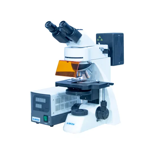
Main Parts of a Fluorescence Microscope
| Part | Main Function |
|---|---|
| Light source | Produces excitation light within the required wavelength range |
| Excitation filter | Selects the wavelength band used to excite the fluorophore |
| Dichroic mirror | Reflects excitation light toward the specimen and transmits much of the emitted fluorescence |
| Objective lens | Focuses excitation light on the sample and collects emitted fluorescence |
| Specimen | Contains fluorescent labels or naturally fluorescent material |
| Emission filter | Blocks residual excitation light and passes the selected emission band |
| Eyepiece | Allows visual observation where the system supports direct viewing |
| Camera or detector | Captures fluorescence images for documentation and analysis |
These components work as a coordinated optical system. Their main task is to deliver the correct excitation light to the sample while isolating the comparatively weak fluorescence signal used to form the image.
How Does a Fluorescence Microscope Work?
- Fluorescent labelling: Where required, the specimen is prepared with a fluorophore that binds to or marks the structure being studied.
- Excitation: The microscope illuminates the specimen with a wavelength band matched to the excitation properties of the fluorophore.
- Fluorescence emission: The excited fluorophore releases longer-wavelength light as it returns to a lower-energy state.
- Optical filtering: The dichroic mirror and emission filter separate useful fluorescence from residual excitation light and unwanted wavelengths.
- Image formation: The filtered fluorescence reaches the eyepiece or camera, forming a high-contrast image of labelled structures.
What Is the Purpose of a Fluorescence Microscope?
The purpose of a fluorescence microscope is to make selected targets within a specimen easier to locate, distinguish, and document. Fluorescent labels can be designed to associate with particular cells, proteins, nucleic-acid sequences, or other structures, depending on the preparation method and research question.
This targeted contrast helps researchers study marked cells, identify microorganisms, locate molecular components, and follow suitable biological processes. Structures that are difficult to distinguish using conventional contrast may become visible when labelled with an appropriate fluorophore and observed using a compatible filter set.
What Is the Main Function of a Fluorescence Microscope?
The main function of a fluorescence microscope is to excite fluorophores within a specimen and collect their emitted light to produce a selective, high-contrast image of labelled targets.
Unlike general bright-field observation, fluorescence imaging can emphasize one or more selected targets rather than displaying all structures with similar contrast. The useful level of detail still depends on the objective, numerical aperture, optical alignment, detector, sample preparation, and imaging method.
Fluorescence Microscope Uses
Cell Biology
Fluorescence microscopy is used to visualize proteins, locate organelles, examine cellular structures, and study suitable dynamic processes through fluorescent markers.
Microbiology
Fluorescent stains and probes can help researchers detect and study bacteria, fungi, and other microorganisms that may be difficult to distinguish using routine transmitted-light observation.
Medical and Diagnostic Research
In tissue and cell studies, fluorescence microscopes support immunofluorescence and related methods used to detect specific targets. Results must be interpreted within the requirements and controls of the applicable laboratory method.
Molecular Biology
Fluorescent probes can be used to study DNA, RNA, and the location of proteins within cells when the probe and preparation method are appropriate for the target.
Pharmaceutical Research
Fluorescence imaging supports suitable cell-based assays, biomarker studies, drug-response research, and other imaging tasks during pharmaceutical development.
Research Laboratories
Research laboratories use fluorescence microscopy for marker-based imaging, experimental documentation, image analysis, and repeatable observation across controlled studies.
Applications of Fluorescence Microscopy
| Field | Example Application |
|---|---|
| Cell biology | Protein and organelle visualization |
| Microbiology | Detection and study of microorganisms |
| Immunology | Immunofluorescence studies |
| Molecular biology | DNA, RNA, and protein localization |
| Pathology research | Controlled tissue and cell analysis |
| Pharmaceutical research | Drug-response and biomarker studies |
| Research laboratories | Advanced fluorescence imaging and documentation |
Across these fields, fluorescence microscopy is valued for its ability to highlight a selected target within a complex specimen. The reliability of that signal depends on appropriate controls, fluorophore selection, filter compatibility, sample preparation, and imaging settings.
Types of Fluorescence Microscopy
Widefield Fluorescence Microscopy
Widefield fluorescence microscopy illuminates and captures the full field of view at once. It is widely used for routine fluorescence observation but may collect out-of-focus light from thicker samples.
Epifluorescence Microscopy
Epifluorescence microscopy uses the same objective to deliver excitation light and collect emitted fluorescence. This is a common optical arrangement in standard fluorescence microscopes.
Confocal Fluorescence Microscopy
Confocal fluorescence microscopy uses optical sectioning to reduce out-of-focus background and produce clearer images of selected planes within suitable specimens.
Total Internal Reflection Fluorescence Microscopy
Total internal reflection fluorescence microscopy, or TIRF, excites a very thin region near the specimen surface. It is useful for studying suitable structures and events close to the coverslip.
Fluorescence Microscope vs Conventional Light Microscope
| Feature | Fluorescence Microscope | Conventional Light Microscope |
|---|---|---|
| Illumination | Selected excitation wavelength band | Visible transmitted or reflected light |
| Image information | Fluorescently labelled or naturally fluorescent targets | General specimen structure and conventional contrast |
| Contrast | Often high for selected targets | Depends on specimen, staining, and contrast method |
| Specificity | Can be highly target-specific with suitable probes | Generally lower target specificity without specialized labelling |
| Fluorescent labels | Often required unless the specimen is naturally fluorescent | Usually not required |
| Common applications | Cell, molecular, microbiological, and advanced research imaging | Routine biological, educational, clinical, and industrial observation |
Advantages of Fluorescence Microscopy
- High specificity for appropriately labelled targets
- Strong contrast between fluorescent targets and background
- Detection of selected molecules or structures within complex samples
- Ability to use multiple compatible fluorophores in suitable multicolour experiments
- Useful for cell, molecular, microbiological, and tissue research
- Compatible with digital imaging, documentation, and analysis workflows
- Supports a wide range of advanced research applications
Limitations of Fluorescence Microscopy
Photobleaching occurs when fluorophores lose their ability to emit light after repeated or prolonged exposure. Phototoxicity can affect living samples when excitation light causes cellular stress or damage during extended imaging.
Autofluorescence and nonspecific background can reduce contrast or be mistaken for a genuine signal. Sample preparation may require additional labelling and controls, while filter sets must match the chosen fluorophores. Fluorescence-ready objectives, illumination systems, cameras, and software can also make the overall system more expensive than a basic light microscope.
Fluorescence Microscope Buying Guide
Application
Define the specimen, target, imaging method, throughput, and level of documentation required.
Fluorophore Compatibility
Confirm that the illumination and filter sets cover the excitation and emission ranges of the fluorophores.
Filter Sets
Match excitation filters, dichroic mirrors, and emission filters to each required fluorescence channel.
Objectives
Compare numerical aperture, optical correction, immersion requirements, field flatness, and transmission characteristics.
Light Source
Assess wavelength coverage, output stability, brightness control, expected lifetime, warm-up needs, and heat generation.
Camera Compatibility
Consider sensitivity, noise, dynamic range, resolution, frame rate, sensor size, and compatibility with the microscope.
Imaging Software
Check acquisition controls, channel handling, measurements, annotations, file formats, analysis, and report export.
Technical Support
Confirm installation, training, calibration, warranty, spare-part availability, and after-sales response.
Choosing for Research Laboratories
The right instrument depends on the required fluorescence channels, specimen type, smallest feature of interest, imaging frequency, and documentation workflow. Filter options, objective quality, camera sensitivity, software support, and upgrade possibilities become especially important when fluorescence imaging is used regularly.
ALMICRO, a brand of Micro Measures & Instruments, is an established Indian microscope manufacturer with a manufacturing background dating to 1978 and a catalogue covering more than 200 microscope configurations. Laboratories comparing a fluorescence microscope manufacturer should evaluate the exact filter sets, objectives, illumination, camera compatibility, software, warranty, and after-sales support offered for their application rather than comparing price alone.
Key Takeaways About Fluorescence Microscopy
- A fluorescence microscope excites fluorophores and detects the longer-wavelength light they emit.
- Excitation and emission filters plus a dichroic mirror separate the useful signal from unwanted light.
- Fluorescence provides selective contrast for labelled cells, proteins, nucleic acids, microorganisms, and other targets.
- Image quality depends on the fluorophore, filter set, objective, detector, specimen preparation, controls, and exposure settings.
- The best microscope configuration is determined by the application and imaging workflow, not maximum magnification alone.
Common Mistakes to Avoid When Using a Fluorescence Microscope
Even a correctly configured fluorescence microscope can produce weak or misleading results when sample preparation, optical settings, or routine care are inconsistent.
- Using the wrong filter set: Filters that do not match the fluorophore's excitation and emission spectra can produce weak signals, spectral overlap, or no useful image.
- Leaving excitation light on too long: Excessive exposure accelerates photobleaching and can increase phototoxicity in live samples.
- Ignoring background fluorescence: Autofluorescence from the specimen, glass, mounting medium, or nonspecific labelling can be mistaken for a true signal. Appropriate controls are essential.
- Skipping objective and immersion-oil care: Dust, fingerprints, dried immersion oil, or incorrect oil can reduce brightness and image clarity and may damage optical surfaces.
- Using an unoptimized fluorophore concentration: Too little label may create a weak signal, while excessive concentration can increase background, nonspecific binding, or quenching.
Practical rule: Match fluorophores and filters carefully, minimize unnecessary exposure, use appropriate experimental controls, and keep the optical path clean.