Research Microscope: Types, Diagram, Uses & Buying Guide in India
Introduction: What Makes a Research Microscope Different?
Most labs do not struggle because their microscope is too weak. They struggle because the image is not sharp. Or the light is uneven. Or the focus slips during a long session. That is the real difference. A research microscope is built for clear images. It gives better detail, steady light and very fine focus. A bigger magnification number on the lens is not the point.
These microscopes are used where the work must be exact and repeatable. Based on the setup, they suit biology, histology, microbiology, clinical work, teaching and imaging.
This guide covers the parts, working, types, uses and buying tips for a microscope for laboratory research.
What Is a Research Microscope?
A research microscope is an optical microscope made for detailed lab work. It focuses on image quality, resolution, sharp focus and controlled light. High magnification alone is not the goal.
One point is often missed. "Research microscope" is not one fixed design. It is a class of quality, not a single model.
The term covers many setups:
- Binocular — for long, easy viewing with both eyes
- Trinocular — has a third port for a camera
- Phase contrast — for clear, unstained samples
- Fluorescence — for samples with dye tags
- Inverted — for cells inside culture flasks
- Digital — built around image capture
So two models can both be called research microscopes. Yet their lenses, light and stage may differ a lot. This is why you should read each spec sheet. Do not trust the label alone.
Research Microscope Diagram and Main Parts
A labelled research microscope diagram is the fastest way to learn the instrument. A research model has more controls than a school microscope. Each one changes the final image.
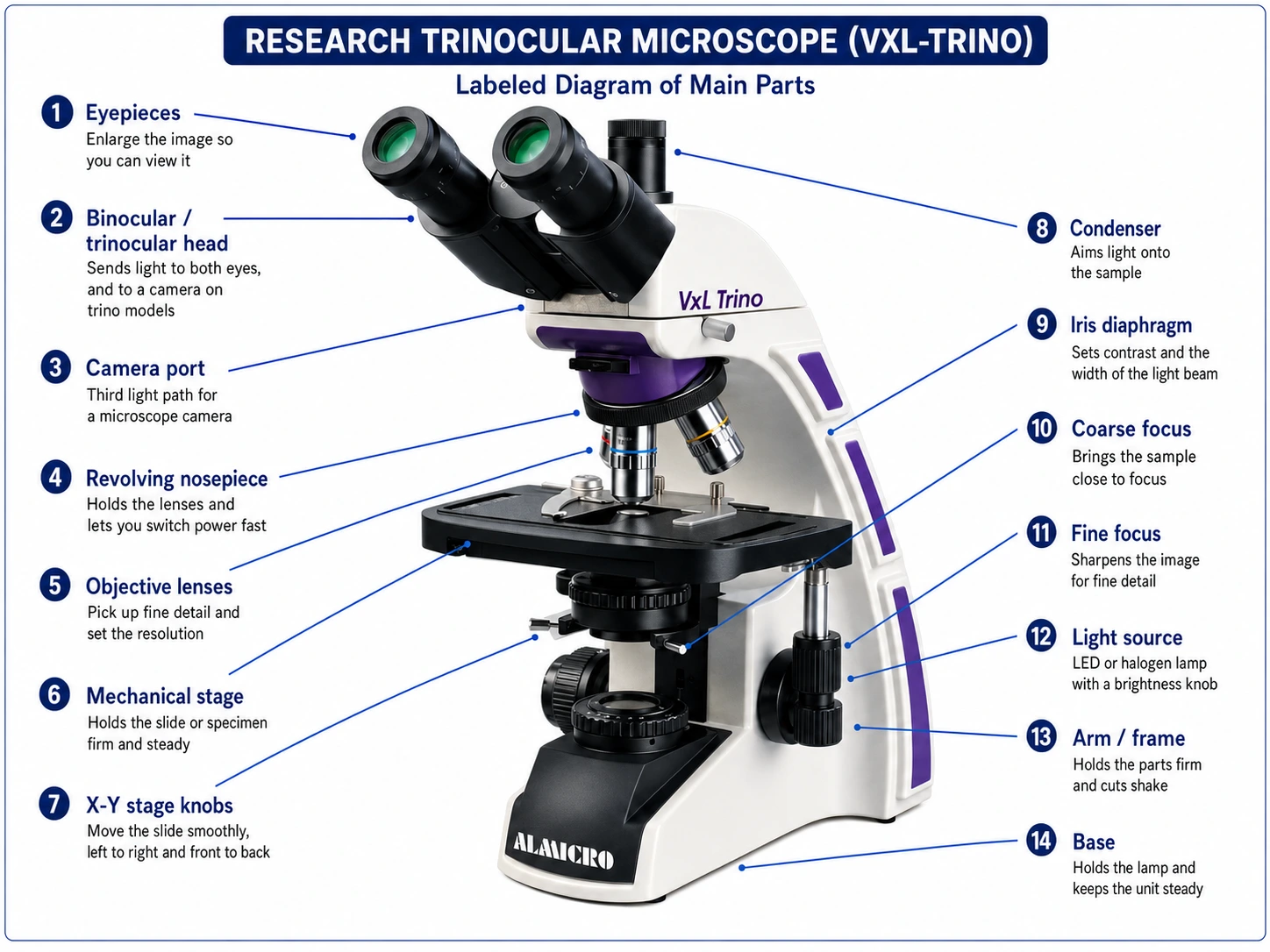
Caption:
Labelled research microscope diagram showing the viewing head, revolving nosepiece, objective lenses, mechanical stage, condenser, iris diaphragm, coarse and fine focus, and the illumination system.
Main parts of a research microscope:
| Part | What It Does |
|---|---|
| Eyepieces | Enlarge the image for viewing |
| Binocular / Trinocular Head | Sends light to both eyes or a camera |
| Camera Port | Provides a light path for a microscope camera |
| Revolving Nosepiece | Holds and switches objective lenses |
| Objective Lenses | Provide magnification and fine detail |
| Mechanical Stage | Holds the slide securely |
| X-Y Stage Knobs | Move the slide smoothly in two directions |
| Condenser | Focuses light onto the specimen |
| Iris Diaphragm | Controls light and image contrast |
| Coarse Focus | Brings the specimen into general focus |
| Fine Focus | Sharpens the image for clear detail |
| Light Source | Provides illumination for the specimen |
| Arm / Frame | Supports the microscope components |
| Base | Supports the microscope and keeps it stable |
How Does a Research Microscope Work?
The light path is simple. Light moves in one order. Each part along the way shapes what you see.
Lamp → condenser → sample → objective lens → viewing head → eyepiece or camera
Here is each step:
- The lamp sends light up through the base.
- The condenser aims that light at the specimen. The iris sets the contrast.
- Light passes through the specimen on the stage.
- The objective lens picks up fine detail and forms a larger image.
- The eyepiece makes that image bigger for your eye.
- On trinocular models, a prism sends part of the light to a camera.
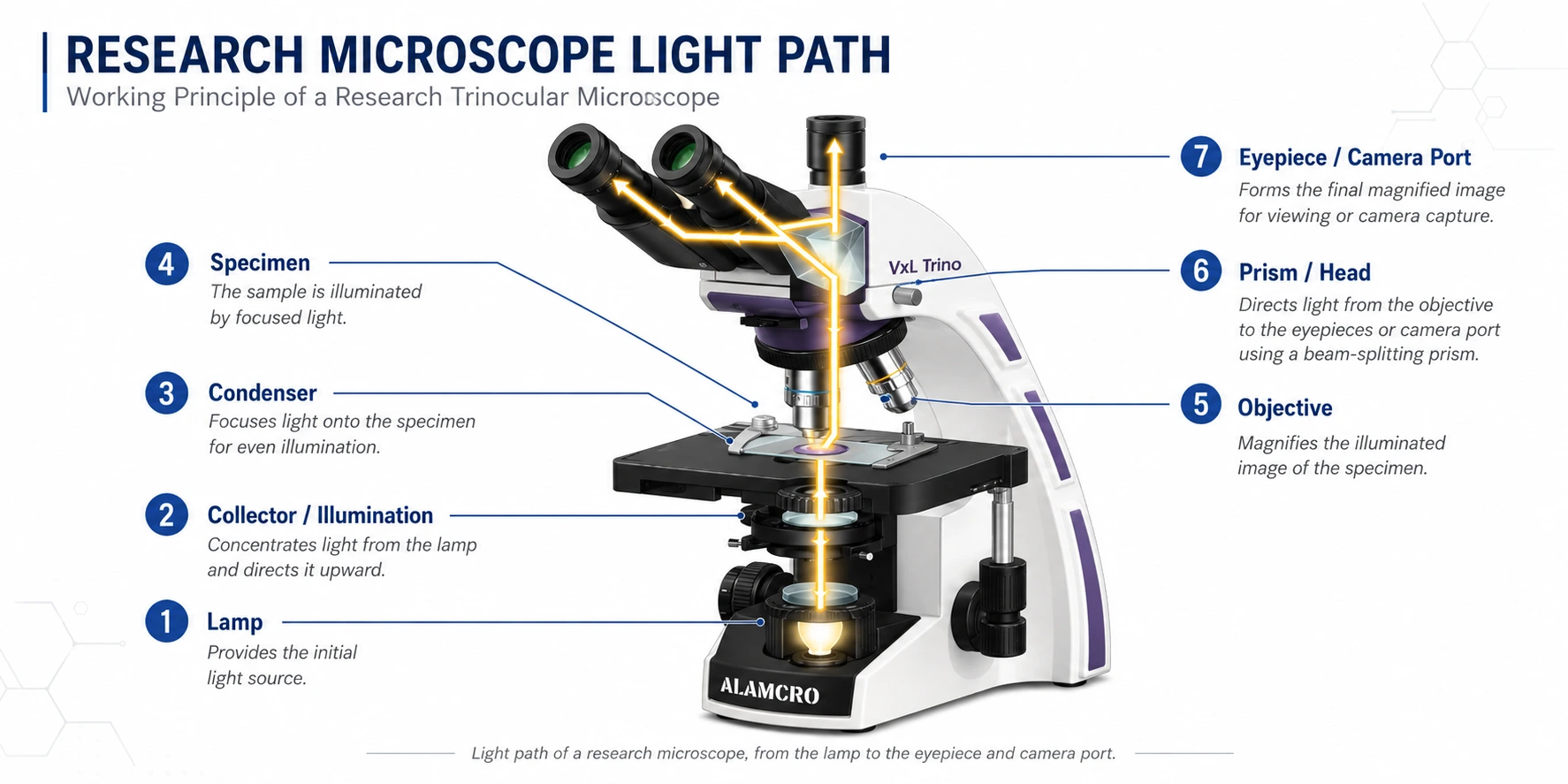
Caption:
How a research microscope works: light travels from the lamp through the condenser and specimen, then through the objective lens to the eyepiece or camera port.
This is why light quality matters so much. If the condenser is off, the image looks flat. Even a great lens cannot fix bad light. Models that support Köhler illumination let you set the light path well. You then get even, glare-free light across the view.
Key Features of a Research Microscope
High-Quality Objective Lenses
The objective lens does most of the work. Research models use achromatic, semi-plan or plan achromatic lenses. Some high-end models use infinity-corrected optics.
Two specs matter more than magnification:
- Numerical aperture (NA) — sets the resolution, or how much fine detail you can see
- Flat field — field flatness decides if the edges stay sharp, not just the middle
A 100x oil immersion objective is common where you need fine cell detail. But not every research model has plan lenses or infinity optics. This changes model to model. Always check the specification sheet.
Precise Coarse and Fine Focusing
Coarse focus brings the sample near focus. Fine focus does the real work at high power.
Coaxial knobs sit on the same axis. They make the job faster and cut hand strain. A tension knob and a focus stop help too. They keep slides and lenses safe.
Mechanical Stage and Specimen Control
A firm stage with smooth X-Y knobs lets you scan a slide in order. You can also come back to the same spot again. That is key when you compare samples or log results.
Illumination
Most new research models use LED light. It stays the same colour, lasts long and runs cool. Some models still use halogen. Brightness control, a good condenser and Köhler setup decide how clear the image looks at high power.
Imaging and Documentation
A trinocular port links the microscope to a camera, monitor or computer. You can then save images, take readings, teach classes and keep records.
If you need images, check camera fit at the time of purchase. Adding it later is often harder.
Types of Research Microscopes
| Research Microscope Type | Best Suited For | Main Advantage |
|---|---|---|
| Binocular Research Microscope | Routine research, long viewing | Easy two-eye viewing |
| Trinocular Research Microscope | Research plus records | Camera can be fitted |
| Phase Contrast Microscope | Clear and live samples | Better contrast, no stain |
| Fluorescence Microscope | Dye-tagged samples | Special fluorescence imaging |
| Inverted Research Microscope | Cells in flasks and dishes | Views from below the vessel |
| Digital Research Microscope | Records and teaching | Direct digital capture |
Here is one point most guides miss. These types are not on the same level.
Binocular vs trinocular is about the viewing head. It tells you how you look, and if a camera can be fitted.
Phase contrast, fluorescence and inverted are about the use. They tell you what kind of sample the model can reveal.
One microscope can be trinocular and phase contrast at the same time. Knowing this saves you from comparing two things that were never rivals.
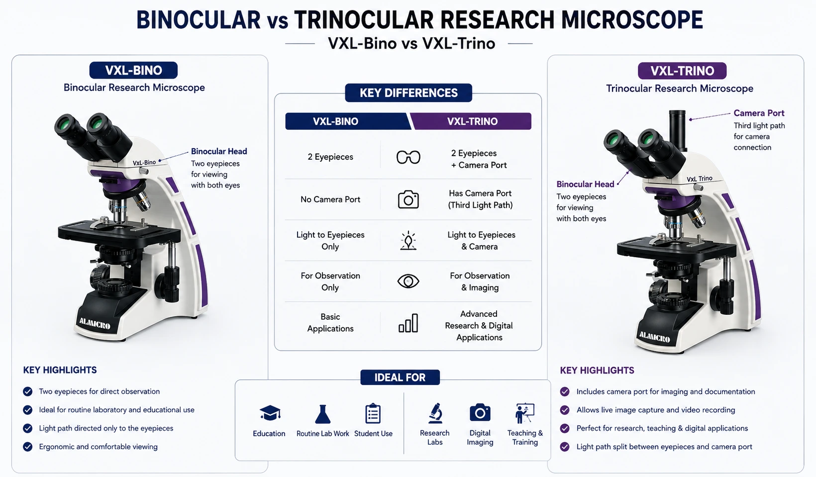
Caption:
Binocular and trinocular research microscope heads compared. The trinocular head adds a third optical port for a microscope camera, allowing image capture and documentation.
Research Microscope Applications
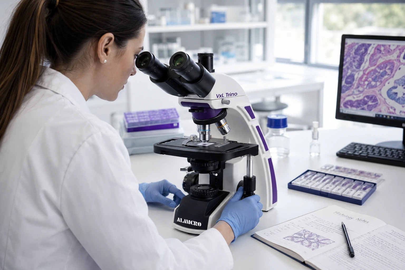
Caption:
A microscope for laboratory research in use for biological samples, tissue sections and prepared slides.
Biological Research
Used to view cells, tissue and prepared samples in teaching and research labs.
Microbiology
Used to study microbes and prepared slides. The method you pick decides if you need brightfield, phase contrast or an oil lens.
Histology and Pathology Research
Used for close study of stained tissue slices. Here, flat field and true colour affect what you read from the slide. Note one thing. Whether a model can be used for clinical tests depends on its approval status. The word "research" on the box does not decide it.
Pharmaceutical and Biotechnology Research
Used for sample checks, product studies and daily lab work that needs steady results.
Universities and Research Institutions
Used for advanced practicals, demos, project work and post-graduate research.
Digital Documentation
Used to capture images for reports, papers, slides and shared class notes.
One rule holds across all of these. Pick a microscope for laboratory research based on the sample and the method. Do not pick it on magnification alone.
Research Microscope vs Standard Laboratory Microscope
| Feature | Standard Lab Microscope | Research Microscope |
|---|---|---|
| Main Use | Routine viewing | Detailed, advanced study |
| Optics | General lab work | Higher optical grade, based on the job |
| Focusing | Coarse and fine | Finer control, often needed |
| Imaging | Optional | Common on advanced models |
| Camera Port | Sometimes | Common on trino models |
| Light Control | Basic to advanced | More control, often preferred |
| Upgrades | Limited, model based | More modular on high-end models |
| Users | Students, routine labs | Researchers, colleges, special labs |
This table shows a trend, not a hard line. Many good lab microscopes work well for research too. The overlap is real.
So do not ask which label a product carries. Ask if the optics, light and stage suit your sample.
How to Choose a Research Microscope for Your Laboratory
Start With the Application
Answer four questions before you compare models:
- What sample will you view?
- Is it live or prepared?
- Which method do you need — brightfield, phase contrast or fluorescence?
- Do you need to save images?
Nearly every other choice flows from these four answers.
Application → Recommended Configuration
Use this table as a quick starting point. It maps common lab applications to the setup that usually suits them best.
| Application | Recommended Configuration | Key Optics & Illumination | Camera Needed |
|---|---|---|---|
| Routine Biology Teaching | Binocular, brightfield | Achromatic 4x–100x, LED | No |
| General Biological Research | Binocular or trinocular, brightfield | Semi-plan or plan achromatic, Köhler | Optional |
| Histology & Tissue Sections | Trinocular, brightfield | Plan achromatic, flat field, Köhler | Yes |
| Microbiology & Bacteria | Binocular or trinocular | 100x oil immersion, high NA | Optional |
| Live or Unstained Cells | Phase contrast | Phase objectives, phase condenser | Optional |
| Cell Culture in Flasks | Inverted | Long working distance objectives | Usually Yes |
| Dye-Tagged Samples | Fluorescence | Fluorescence filters, dedicated light source | Yes |
| Image Records & Teaching Demos | Trinocular, digital | Camera port with matched adapter | Yes |
| Pharma & QC Labs | Trinocular, brightfield | Plan achromatic, steady LED | Yes |
Two notes before you use this table.
First, the rows are starting points, not rules. Your sample and method still decide the final call.
Second, some rows can be combined. A trinocular head can also carry phase contrast optics. Head type and application type are separate choices.
Evaluate Optical Quality, Not Magnification Alone
Check the objective type, numerical aperture, resolution, field flatness and contrast. A 1000x image from a weak objective is just a bigger blur. High magnification with low resolution adds nothing.
Choose Binocular or Trinocular
Pick binoculars if you only need to view. It costs less. Pick trinocular if you need a camera now, or plan to add one later.
Check Illumination
Check the lamp type, brightness control and condenser. If you need even light across the whole view, choose a model that supports Köhler illumination.
Consider Ergonomics
For long sessions, check the head angle and eye-width setting. Also check where the stage and focus knobs sit. Comfort affects your data. A tired eye misses detail.
Check Camera and Upgrade Compatibility
Check the camera port size, adapters and software. Ask if you can add lenses, a condenser or a new head later.
Evaluate Service and Spare Parts
For colleges and labs, this often decides the deal. Check the warranty, service time and spare part supply. Ask if setup and training are included.
Research Microscope in India: What Should Buyers Look For?
Buyers looking for a research microscope in India should fix the use case first. Then decide the optical setup. Only after that should you compare brands or prices.
Doing it the other way is the main cause of a bad buy.
Use this checklist while you shortlist:
- Use case and sample type
- Full optical specification, not just the magnification range
- Lens set, including an oil lens if needed
- Lamp type, brightness control and condenser
- Binocular or trinocular head
- Camera and software fit
- Warranty terms
- Support and setup help
- Spare part supply over the years
- Ability to handle bulk or tender orders
Many labs in India buy through tenders. Papers matter there. Ask for a printed spec sheet. You can then check the unit you receive against the quote.
Choosing a Research Microscope Manufacturer in India
The product and the supplier are two separate calls. The second one decides how well the first works over the next ten years.
Judge a research microscope manufacturer in India on these points:
- Know-how — can they suggest a setup for your sample, or only quote a price?
- Range — do they have many research models, or push one model for every job?
- Options — lens types, condensers and lamp choices
- Making capacity — do they build in-house, or only trade?
- Papers — clear and full specification sheets
- Custom builds — can they tune a model to your need?
- Quality checks — testing before dispatch
- After-sales — how fast do they respond?
- Spare parts — long-term supply of lamps, lenses and gears
- Bulk supply — past work with tenders and college orders
Here is one example of how such firms are set up. Micro Measures & Instruments began in 1978. It is the company behind the ALMICRO microscope brand. Its range covers school, lab, research, digital, industrial and special models. Note the split between the company name and the brand name. Both may show up on bills, catalogues and tender papers.
What Should You Expect From a Leading Microscope Manufacturer in India?
A good microscope manufacturer in India should give you:
- Correct specifications you can verify
- Advice based on your use case, not a stock quote
- More than one optical option
- Full brochures and product papers
- Clear warranty and support terms
- Spare parts for years to come
- Proof of bulk and tender supply
- Help after the unit is installed
If a supplier will not answer specification questions in writing, treat that as an early warning.
For a closer look at how to judge a supplier, read our guide on picking a Leading Microscope Manufacturer in India. Our Different Types of Microscopes guide also shows where research models sit in the wider family.
ALMICRO Research Microscope Options
If you are still comparing setups, these four cover most research needs:
- Research Binocular Microscope — for long viewing in teaching and research labs
- Research Trinocular Microscope — adds a camera port for images and records
- Advanced Research Microscope — for jobs that need better optics and finer light control
- Research Microscope with Digital Imaging — built for capture, display and records
Current models include the BM-9bi and BM-8tr series. The range also has VXL-Bino, VXL-Trino and EXL units. Each has its own optics and illumination specification. So check each specification sheet against your use case before you decide.
Research Microscope: Questions Buyers Ask Most
What is a research microscope?
A research microscope is an optical microscope made for detailed lab work. It puts image quality, resolution, sharp focus and steady light above high magnification. It may be a binocular, trinocular, phase contrast, fluorescence, inverted or digital model, based on the job.
Which microscope is commonly used for laboratory research?
It depends on the sample and method. For light-based biology work, compound binocular and trinocular models are the usual choice. Live cell work in flasks needs an inverted model. Clear, unstained samples show up better with phase contrast.
What is the difference between a research microscope and a laboratory microscope?
The two overlap a lot. Research models often have better optics, finer focus, more illumination control and a camera port. But many good lab microscopes handle research work well. Compare specs, not labels.
Is a binocular or trinocular microscope better for research?
Pick trinocular if you need to save images, keep records or show the view on a screen. If you only need to look, a binocular model gives the same view for less money.
What parts should be included in a research microscope diagram?
A full diagram should label the eyepieces, head, camera port, nosepiece, lenses, stage and X-Y knobs, condenser, iris, coarse and fine focus, lamp, arm and base.
How do I choose a research microscope in India?
Work in this order: use case, then optics, then light, then imaging, then service. Fix the sample and method first. That stops you from paying for extras you will never use.
Key Takeaways Before You Choose a Research Microscope
The right research microscope is set by the work it must do. It is not set by the highest magnification on the box. Once you know the sample and method, the correct setup is usually clear.
Focus on five things. Objective quality and numerical aperture. Illumination control. Focus accuracy. A steady stage. And whether you need to save images. These shape your image far more than magnification does.
Give the supplier equal weight. Warranty, spare parts and real support decide if the unit still works well years from now.