What Is a Research Microscope?
There is not one single “research microscope” technique. The right configuration depends on the specimen being examined and the information the laboratory needs to obtain. A cytology laboratory preparing stained slides has very different equipment requirements from a cell-culture facility monitoring live, unstained cells, even though both operate within research microscopy.
Research-grade performance is not defined by magnification alone. It results from a combination of optical and mechanical choices matched to the specimen and intended output, whether that output is visual assessment, photomicrography or quantitative measurement.
The parameters that determine whether a microscope is suitable for research use include:
- Infinity-corrected optics
- Objective type and numerical aperture
- Illumination system
- Contrast technique
- Mechanical stage and focusing
- Trinocular imaging port
- Camera and software compatibility
Key principle: Optical resolution depends strongly on illumination wavelength and numerical aperture, not on maximum magnification alone. This distinction is essential when comparing research microscope specifications.
Key Features of a High-End Research Microscope
A well-specified research microscope commonly includes:
- Infinity-corrected optical system
- Plan achromatic or higher-correction objectives
- High numerical-aperture objectives where the application requires them
- Trinocular observation and imaging head
- Köhler illumination capability
- LED or another application-appropriate illumination source
- Coaxial coarse and fine focusing
- Precision mechanical X-Y stage
- Brightfield and compatible contrast techniques
- Digital camera and imaging-software compatibility
Important: Not every feature is mandatory for every research application. A histology laboratory examining stained brightfield slides has different priorities from a cell-biology laboratory performing live-cell fluorescence imaging. Use this as an application-based checklist, not a universal pass-or-fail specification.
Research Microscope Optical System
Infinity-Corrected Optical System
In an infinity-corrected microscope, light emerging from the objective travels as an approximately parallel, collimated beam through an “infinity space” before a separate tube lens forms the intermediate image. Technical descriptions of infinity optics explain how this optical space allows compatible filters, beam splitters and other components to be introduced into the light path with controlled effects on focus.
The practical benefit is modularity. It can make it easier to integrate components such as fluorescence filter cubes, polarisers and camera ports within a compatible optical system.
Infinity correction does not automatically guarantee greater resolution than every finite-conjugate microscope. Resolution is principally governed by factors including objective numerical aperture and illumination wavelength.
Plan Achromatic Objectives and Optical Correction
Objective terminology is often used loosely, so the main classes should be distinguished carefully:
- Achromatic objectives: Correct chromatic aberration for selected wavelengths and provide suitable performance for routine observation.
- Plan objectives: Improve field flatness so more of the image remains in focus from the centre toward the edge.
- Plan achromatic objectives: Combine chromatic correction with improved flat-field performance, making them suitable for general research imaging and photomicrography.
Higher-correction classes, such as plan fluorite and plan apochromatic objectives, extend optical correction and may be valuable for demanding fluorescence or quantitative imaging. However, the word “Plan” does not itself indicate greater resolving power. Field flatness and optical resolution are separate properties.
| Objective Type | Chromatic Correction | Field Flatness | Typical Use |
|---|---|---|---|
| Achromatic | Basic correction | Limited | Routine viewing |
| Plan Achromatic | Basic correction | Corrected | Research imaging and photomicrography |
| Plan Fluorite / Semi-Apo | Improved correction | Corrected | Fluorescence and higher-precision imaging |
| Plan Apochromatic | Highest correction class | Corrected | Demanding research and quantitative imaging |
Numerical Aperture and Resolution: Specifications Buyers Should Check
Why Numerical Aperture Matters
Numerical aperture (NA) describes the range of light-gathering angles an objective can accept from the specimen within a particular medium, such as air, water, glycerine or immersion oil. It is one of the principal variables that determines the resolution limit of an optical system.
Abbe’s resolution relationship shows that resolvable distance is approximately proportional to wavelength divided by twice the numerical aperture. In practical terms, a higher-NA objective or shorter-wavelength illumination can resolve finer detail, up to the diffraction limit of the optical system.
Higher numerical aperture involves trade-offs. It commonly produces a shallower depth of field and a shorter working distance, which may matter when examining thick specimens or manipulating samples beneath the objective.
Magnification vs Useful Resolution
Common objective magnifications include 4X, 10X, 20X, 40X and 100X. A claim such as “up to 1000X magnification” does not, by itself, demonstrate research-grade image quality.
Once an optical system reaches its diffraction-limited resolving power, additional magnification only enlarges the existing image. It does not add new spatial information and may result in empty magnification.
Buyer takeaway: Do not compare research microscopes by maximum magnification alone. Compare objective numerical aperture, optical correction, contrast performance and compatibility with the intended specimen.
Trinocular Head and Camera Integration
A binocular head directs the image to two eyepieces. A trinocular head adds a third port, normally connected through a beam splitter, which directs some or all of the available light toward a camera.
The third port supports:
- Image documentation and archiving
- Video capture
- Quantitative measurement workflows
- Image sharing across research and teaching teams
Camera integration also requires evaluation of adapter magnification, sensor size in relation to the field of view, pixel sampling, exposure control, image-acquisition software and compatibility with the microscope’s photo port.
A trinocular head does not improve optical resolution by itself. It changes how the image can be accessed and recorded; the objective and optical system still determine the resolved specimen detail.
| Feature | Binocular | Trinocular |
|---|---|---|
| Eyepiece viewing | Yes | Yes |
| Dedicated camera port | No | Yes |
| Documentation workflow | Limited or adapter-dependent | Integrated |
| Optical resolution | Determined by objective and NA | Determined by objective and NA |
Research Microscope Illumination System
Köhler Illumination
Köhler illumination is widely used to produce even, controlled specimen illumination in research microscopy. It places an image of the light source at the condenser aperture plane rather than at the specimen plane, helping prevent the source structure from appearing in the final image.
The two main controls are the field diaphragm, which limits the illuminated specimen area, and the condenser aperture diaphragm, which controls the angle and effective numerical aperture of the illuminating cone. Evident’s Köhler illumination guide explains the role of condenser adjustment in achieving consistent microscope illumination.
Köhler illumination is an illumination-control method, not an independent resolution-enhancing technology. It improves illumination uniformity and contrast control, while the diffraction-limited resolution remains governed primarily by NA and wavelength.
LED vs Halogen Illumination
| Parameter | LED | Halogen |
|---|---|---|
| Heat generation | Generally lower | Generally higher |
| Energy use | Generally lower | Higher |
| Service life | Generally longer | Generally shorter |
| Spectrum | Depends on LED design | Broad and continuous |
| Best suited for | Application-dependent | Application-dependent |
LED sources usually operate with lower heat and energy consumption, which can be useful during long observation sessions and when examining heat-sensitive live specimens. However, LED illumination does not automatically produce a better image. Image quality depends on the source spectrum, optical coupling, specimen, detector and alignment of the complete illumination system.
Mechanical Stage and Coaxial Focusing System
The mechanical system receives less attention than the optics, but it strongly affects day-to-day usability and repeatability. Important features include:
Coaxial coarse and fine focus controls positioned for efficient one-handed operation
Fine adjustment for precise focal positioning, particularly at high NA where depth of field is shallow
A mechanical X-Y stage for controlled and repeatable specimen movement
A stage travel range compatible with the intended slide or vessel format
Focus-tension adjustment where available
Stage repeatability is particularly useful in imaging and multi-field workflows where the operator may need to return to the same coordinates.
Specification caution: Do not accept a precise mechanical-accuracy claim, such as a one-micron focusing figure, unless it is confirmed in the documentation for the exact microscope model. There is no universal focusing specification that applies to every instrument marketed as research grade.
Contrast Techniques Used in Research Microscopy
| Technique | Best Suited For |
|---|---|
| Brightfield | Stained or naturally high-contrast specimens |
| Darkfield | Specimens where scattered light provides useful contrast |
| Phase Contrast | Transparent, unstained specimens, particularly live cultured cells |
| Fluorescence | Fluorescently labelled structures and molecularly specific imaging |
Each technique uses a different image-formation principle. Brightfield commonly relies on absorption or staining, phase contrast converts phase differences associated with refractive-index and thickness variations into intensity contrast, fluorescence detects light emitted by fluorophores, and darkfield forms an image primarily from scattered light.
Select the contrast method according to the specimen and the information required, rather than choosing the technique that appears most advanced on a specification sheet.
Types of Research Microscopes and Their Applications
| Research Microscope Type | Primary Application |
|---|---|
| Upright Brightfield | Histology and stained slides |
| Phase Contrast | Live and unstained cells |
| Fluorescence | Label-specific biological imaging |
| Inverted | Cell and tissue culture |
| Polarizing | Birefringent materials |
| Metallurgical / Reflected Light | Opaque materials and surfaces |
| Digital / Camera-Integrated | Documentation and quantitative imaging |
Research Microscope Specifications: What Should Buyers Compare?
| Specification | What to Evaluate |
|---|---|
| Optical system | Infinity-corrected or other intended optical configuration |
| Objectives | Plan achromatic or higher correction according to the application |
| Numerical aperture | Application-specific resolving requirement |
| Head | Binocular or trinocular configuration |
| Eyepiece | Field number and viewing comfort |
| Illumination | LED, halogen or specialised source |
| Illumination control | Köhler capability and condenser system |
| Condenser | NA and contrast-method compatibility |
| Stage | X-Y travel and specimen compatibility |
| Focusing | Coarse and fine coaxial controls |
| Contrast | Brightfield, phase contrast, darkfield, fluorescence or other required method |
| Camera port | Adapter compatibility and light-path configuration |
| Camera | Sensor, sampling and acquisition requirements |
| Software | Capture, calibration, measurement and analysis features |
How to Select a Research Microscope for Your Laboratory
Define the specimen: Determine whether it is fixed or live, stained or unstained, transparent or opaque, and whether it is held on a slide, in a culture vessel or as bulk material.
Identify the required contrast method: Select brightfield, phase contrast, fluorescence, darkfield, polarization or reflected light according to the specimen and required information.
Match objective NA and correction: Begin with the resolving requirement rather than the highest magnification printed on the brochure.
Choose an upright or inverted configuration: Base the decision on sample geometry and workflow. Inverted systems are commonly used for cell and tissue cultures held in vessels.
Define documentation requirements: If digital imaging is necessary, evaluate the trinocular port, adapter, camera sensor and software as one connected system.
Evaluate illumination: Check stability, spectral output, heat generation and compatibility with the selected contrast technique.
Review mechanical and ergonomic requirements: Consider focusing controls, stage position, viewing angle and control accessibility. These factors affect comfort during prolonged laboratory use.
Research Microscope Features by Laboratory Requirement
| Laboratory Requirement | Priority Feature |
|---|---|
| Routine stained-slide research | Brightfield with plan objectives |
| Live-cell observation | Phase contrast matched to specimen geometry |
| Cell culture | Inverted configuration |
| Molecular localisation | Fluorescence |
| Image documentation | Trinocular port and compatible camera |
| Quantitative imaging | Correct optics with calibrated camera and software |
| Long-duration observation | Ergonomic controls and stable illumination |
| Material-surface analysis | Reflected-light or metallurgical configuration |
The correct question is not “Which research microscope is best overall?” but “Which configuration fits the laboratory’s actual specimen and workflow?” There is no universally best research microscope—only a better or worse application fit.
Common Mistakes When Comparing Research Microscopes
Comparing only maximum magnification: Resolution depends on numerical aperture and wavelength, not only the largest magnification number.
Assuming trinocular means better resolution: A trinocular head changes imaging access and camera integration, not the objective’s resolving power.
Ignoring numerical aperture: NA directly influences resolution; magnification and correction class cannot substitute for it.
Assuming more camera megapixels always mean greater detail: Optical resolution and detector sampling are separate limits. A high-megapixel sensor cannot recover detail the objective did not resolve.
Selecting contrast without considering the specimen: Each technique uses a different image-formation mechanism, and an unsuitable method can obscure the information being investigated.
Research Microscope Diagram
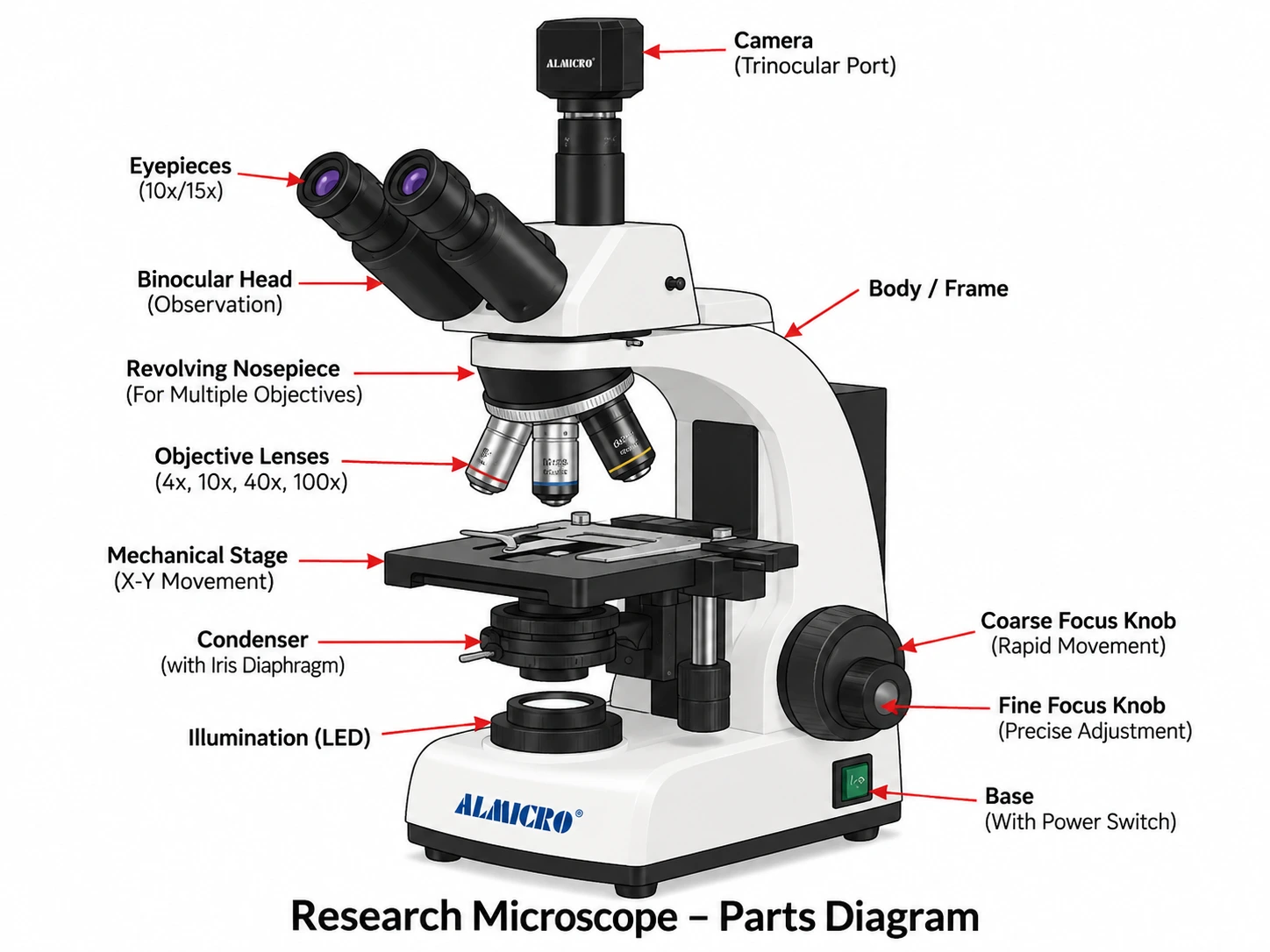
Recommended visual: Use a labelled trinocular research microscope rather than a basic school microscope.
The diagram should identify the eyepieces, trinocular or photo port, observation head, revolving nosepiece, plan objective lenses, mechanical stage, X-Y stage controls, condenser, aperture diaphragm, field diaphragm, coaxial coarse and fine focus controls, LED illuminator, arm or frame, and base.
Final Selection Considerations for Research Laboratories
A research microscope should be selected according to the specimen, required optical resolution, contrast technique, objective correction, illumination, mechanical precision and intended imaging workflow rather than maximum magnification alone.
Infinity-corrected optics, plan objectives, controlled illumination and camera integration can all be valuable, but the importance of each feature depends on the research application. Treat the specification sheet as one integrated optical and imaging system instead of a collection of isolated headline features.
ALMICRO has manufactured microscopes and laboratory instruments in Ambala Cantt, Haryana, since 1978. Its current range includes more than 200 microscope models covering brightfield, phase contrast, fluorescence, inverted and metallurgical configurations for research, educational and industrial laboratories.
Frequently Asked Questions
What Specifications Should I Check Before Selecting a Research Microscope?
Check the optical system, objective correction, numerical aperture, head configuration, illumination, condenser, mechanical stage, focusing system, contrast compatibility and camera integration. No single specification tells the complete story; the components must be evaluated together against the specimen and workflow.
Is an Infinity-Corrected Microscope Better for Research?
Infinity-corrected systems provide an optical space that can make it easier to integrate compatible filters, beam splitters and other components. This is a practical advantage, but it does not independently guarantee better resolution or image quality. Objective NA, correction, illumination and system alignment remain important.
Is a Trinocular Microscope Necessary for Laboratory Research?
Not necessarily. A trinocular head is useful when the workflow requires camera-based documentation, recording or digital analysis. It does not provide greater optical resolution than a binocular configuration using the same objectives and optical system.
Are Plan Achromatic Objectives Suitable for Research Microscopy?
Yes. They are suitable for many general research-imaging applications where chromatic correction and field flatness are important. More highly corrected objectives may be preferable for demanding fluorescence or quantitative work, depending on the required NA, correction, working distance and imaging method.