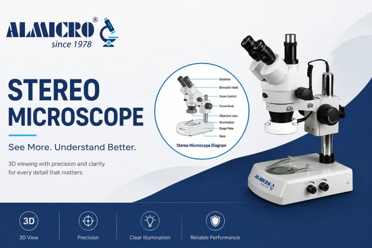
Stereo Microscope – Principle, Parts, Diagram, Uses & Applications
Stereo microscopes are advanced optical instruments used for 3D viewing of larger specimens in laboratories, industries, education, and research. Unlike compound microscopes, they provide depth perception, wide viewing area, and comfortable specimen handling, making them ideal for electronics inspection, biological dissection, jewelry analysis, forensic science, and industrial quality control.
What is a stereo microscope?
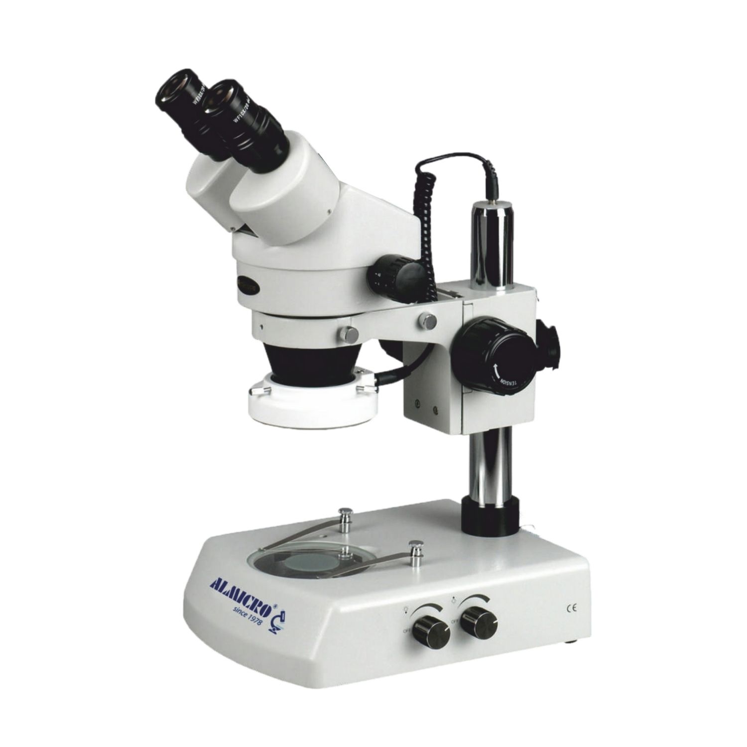
A stereo microscope, also called a dissecting microscope, is a low-magnification optical microscope used for viewing specimens in three dimensions. To understand what a stereo microscope is, it uses two separate optical paths to create a realistic 3D image with depth perception. Stereo microscopes are widely used for observing biological samples, electronics, insects, plants, jewelry, and industrial components with comfortable handling and wide viewing area.
Key characteristics include:
- Three-dimensional image formation
- Long working distance
- Wide viewing field
- Comfortable binocular viewing
- Real-time specimen handling
- Surface structure observation
Stereo microscopes are commonly used in:
- Educational laboratories
- PCB inspection
- Electronics repair
- Biological dissection
- Jewelry examination
- Medical training
- Industrial quality control
The stereo microscope function focuses more on external morphology and structural observation rather than cellular-level magnification.
Stereo Microscope Diagram
Understanding the stereo microscope diagram is essential for learning how the instrument works. Each component contributes to image formation, magnification control, illumination, and specimen stability.
Main Components in a Stereo Microscope Diagram
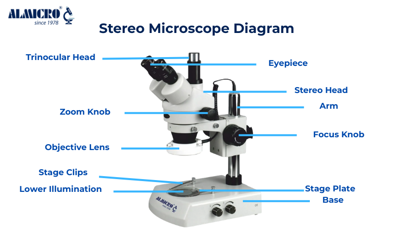
| Part | Function |
|---|---|
| Eyepiece | Magnifies the image for viewing |
| Objective Lens | Produces primary magnification |
| Binocular Head | Provides dual optical viewing paths |
| Zoom Knob | Adjusts magnification smoothly |
| Focus Knob | Moves microscope vertically for focusing |
| Stage Plate | Holds specimen |
| Stage Clips | Secure sample in position |
| Illumination System | Provides reflected or transmitted light |
| Arm | Supports optical head |
| Base | Stabilizes the microscope |
Optical Path Explanation
The stereo microscope diagram usually shows two independent optical channels angled toward the specimen. Each eye receives a slightly different image, which the brain combines into a stereoscopic 3D image.
This dual optical configuration is the core of the stereo microscope working principle.
Illumination System
Stereo microscopes generally use:
- Top reflected illumination
- Bottom transmitted illumination
- LED ring lights
- Fiber optic lighting systems
These lighting systems enhance contrast and surface visibility.
Parts of Stereo Microscope
Understanding the individual stereo microscope parts helps users operate the system effectively and maintain accurate observation quality.
Eyepiece
The eyepiece, also called the ocular lens, is positioned at the top of the microscope. It further magnifies the image produced by the objective lens.
Typical eyepiece magnifications include:
- 10x
- 15x
- 20x
Modern stereo microscope for laboratory applications may include wide-field eyepieces for greater viewing comfort.
Objective Lens
The objective lens creates the primary magnified image of the specimen. Stereo microscopes use low-power objectives because they are intended for larger objects and surface observation.
Common objective ranges include:
- 0.5x
- 1x
- 2x
- Zoom objective systems
Binocular Head
The binocular head contains two separate optical paths. This is the defining feature that enables true depth perception.
Benefits include:
- Reduced eye strain
- Better depth interpretation
- Comfortable long-term viewing
Stage Plate
The stage plate supports the specimen during observation. Stereo microscopes may use:
- Black/white reversible plates
- Frosted glass plates
- Transparent stage plates
Stage Clips
Stage clips secure specimens in place during analysis or manipulation.
These are especially useful for:
- Dissection work
- PCB inspection
- Jewelry examination
Illumination System
The illumination system plays a critical role in image quality.
Common lighting systems include:
- LED illumination
- Halogen lighting
- Ring lights
- Fiber optic systems
Industrial stereo microscope systems often use adjustable directional lighting for reflective surfaces.
Zoom Control
The zoom knob allows smooth magnification changes without replacing lenses.
Stereo zoom microscope systems are preferred because they provide:
- Continuous magnification adjustment
- Better workflow efficiency
- Improved inspection flexibility
Focus Knob
- The focus knob moves the optical head vertically to sharpen the image.
- Coarse focusing is generally sufficient because stereo microscopes use lower magnification.
Arm and Base
- The arm supports the optical assembly while the base stabilizes the entire microscope system.
- Heavy-duty industrial microscopes often use reinforced bases for vibration reduction.
Stereo Microscope Principle
The stereo microscope principle is based on binocular stereoscopic vision using two independent optical paths. Unlike compound microscopes that create a flat 2D image, stereo microscopes produce a three-dimensional visual effect because each eye receives a slightly different viewing angle.
Core Stereo Microscope Working Principle
The stereo microscope working principle involves:
- Illumination of the specimen
- Reflection or transmission of light
- Entry of light into separate optical paths
- Independent image formation for each eye
- Brain interpretation of depth differences
This process creates natural depth perception.
Separate Optical Channels
Each optical path contains:
- Objective lens
- Prism system
- Eyepiece
The slight angular difference between left and right optical channels mimics human binocular vision.
Stereoscopic Image Formation
Because both eyes receive different perspectives, the brain reconstructs the image with depth information.
This enables users to:
- Observe surface texture
- Manipulate specimens
- Perform micro-assembly tasks
- Conduct dissections accurately
Magnification System
Stereo microscopes use low-power optical systems with wide working distance.
Typical stereo microscope magnification ranges include:
| Type | Magnification |
|---|---|
| Basic Educational | 10x–20x |
| Laboratory Models | 20x–40x |
| Stereo Zoom Microscope | 5x–80x |
| Industrial Models | Up to 100x |
How Does a Stereo Microscope Work?
The working process of a stereo microscope is relatively straightforward but highly effective for practical observation.
Step 1: Illumination
Light is directed toward the specimen using:
- Reflected top light
- Transmitted bottom light
Reflective illumination is most common for solid objects.
Step 2: Light Reflection from Specimen
- The specimen reflects light into the objective lenses.
- Surface texture and structure become visible during this stage.
Step 3: Objective Lens Magnification
- The objective lenses create the initial magnified image.
- Because stereo microscopes use low magnification, they maintain a large field of view.
Step 4: Separate Image Formation
- Each optical path forms an independent image.
- This is the foundation of the stereo microscope principle.
Step 5: Brain Depth Interpretation
The brain merges both images into a realistic 3D image.
This provides:
- Spatial orientation
- Accurate specimen handling
- Improved hand-eye coordination
For example, during PCB soldering inspection, technicians can clearly observe solder joints, component placement, and surface defects in three dimensions.
Stereo Microscope Magnification
Stereo microscope magnification is intentionally lower than compound microscopes because the goal is surface observation rather than cellular analysis.
Typical Magnification Range
Most stereo microscopes operate between:
- 5x to 80x
Some industrial systems may exceed this range using auxiliary lenses and digital enhancement.
Working Distance
A major advantage is long working distance.
This allows users to:
- Handle tools under the microscope
- Perform dissections
- Repair components
- Conduct assembly work
Magnification Comparison Table
| Microscope Type | Magnification Range | Image Type |
|---|---|---|
| Stereo Microscope | 5x–80x | 3D |
| Compound Microscope | 40x–1000x | 2D |
| Digital Microscope | Variable | Digital |
| Industrial Inspection Microscope | 10x–200x | 3D/Hybrid |
Stereo Microscope Uses
The section on stereo microscope uses is one of the most important because these microscopes are widely used across numerous industries.
Electronics Inspection
Stereo microscopes are heavily used for:
- PCB inspection
- Solder joint analysis
- Semiconductor inspection
- Microelectronics assembly
The 3D viewing capability helps technicians identify defects accurately.
Jewelry Inspection
Jewelers use stereo microscopes for:
- Gemstone grading
- Surface inspection
- Engraving analysis
- Stone setting
Depth perception is critical during precision handling.
Biological Dissection
As a dissecting microscope, it is widely used in biology laboratories.
Applications include:
- Insect dissection
- Plant structure analysis
- Tissue examination
- Educational demonstrations
Educational Laboratories
The uses of stereo microscope in schools and universities are extensive.
Students can observe:
- Leaves
- Insects
- Seeds
- Small organisms
- Surface structures
The wide field of view makes learning easier.
Medical Training
Medical institutions use stereo microscopes for:
- Surgical training
- Dental procedures
- Tissue handling
- Microinstrument practice
Entomology
Insect researchers use stereo microscopes for:
- Species identification
- Morphological analysis
- Wing structure observation
Botanical Studies
Stereo microscope in biology laboratories supports:
- Seed analysis
- Flower structure examination
- Plant pathology
Industrial Quality Control
Industrial stereo microscope systems help manufacturers inspect:
- Machined parts
- Surface coatings
- Weld joints
- Precision components
Watch Repair
Watchmakers rely on stereo microscopes for:
- Gear inspection
- Precision assembly
- Micro-repair tasks
Surgical Applications
Advanced medical stereo microscopes assist surgeons during:
- Microsurgery
- ENT procedures
- Ophthalmic operations
The combination of magnification and depth perception is highly valuable.
Applications of Stereo Microscope in Different Industries
Medical Industry
Applications include:
- Tissue handling
- Surgical training
- Dental inspection
- Clinical observation
Research Laboratories
Researchers use stereo microscopes for:
- Specimen preparation
- Surface analysis
- Biological observation
Industrial Manufacturing
Industrial microscope systems support:
- Quality inspection
- Assembly verification
- Defect analysis
Semiconductor Industry
Used for:
- Chip inspection
- Circuit analysis
- Bond wire inspection
Forensic Science
Forensic investigators use stereo microscopes to analyze:
- Hair samples
- Fibers
- Tool marks
- Surface evidence
Agriculture
Applications include:
- Seed quality analysis
- Pest identification
- Plant disease observation
Textile Industry
Stereo microscopes help examine:
- Fiber structure
- Fabric defects
- Weaving quality
Gemology
Gemologists inspect:
- Gem inclusions
- Surface finish
- Authenticity indicators
Industry Applications Table
| Industry | Application |
|---|---|
| Electronics | PCB inspection |
| Biology | Dissection |
| Medical | Surgical training |
| Gemology | Gem analysis |
| Textile | Fiber inspection |
| Agriculture | Pest identification |
| Forensics | Trace evidence analysis |
| Manufacturing | Quality control |
Stereo Microscope vs Compound Microscope
Understanding the difference between these microscopes is important for selecting the correct instrument.
| Feature | Stereo Microscope | Compound Microscope |
|---|---|---|
| Image Type | 3D | 2D |
| Magnification | Low | High |
| Working Distance | Long | Short |
| Illumination | Reflected | Transmitted |
| Specimen Type | Solid objects | Thin slides |
| Depth Perception | Excellent | Minimal |
| Applications | Inspection/dissection | Cellular analysis |
| Field of View | Wide | Narrow |
Advantages of Stereo Microscope
3D Imaging
Provides natural depth perception.
Long Working Distance
Allows specimen handling during observation.
Real-Time Observation
Useful for dynamic manipulation tasks.
Wide Field of View
Large specimens can be observed comfortably.
Better Hand-Eye Coordination
Critical for repair and dissection work.
Comfortable Viewing
Binocular viewing reduces eye strain.
Limitations of Stereo Microscope
Despite their advantages, stereo microscopes also have limitations.
Lower Magnification
They cannot visualize cellular organelles like compound microscopes.
Limited Cellular Observation
Not suitable for microbiology requiring ultra-high magnification.
Higher Cost for Advanced Models
Industrial and digital systems may be expensive.
Optical Constraints
Resolution is lower compared to compound microscope systems.
How to Choose the Best Stereo Microscope
Selecting the right stereo microscope depends on intended applications.
Magnification Range
Choose according to specimen size and detail requirements.
Lighting System
LED lighting is preferred for:
- Energy efficiency
- Uniform illumination
- Low heat generation
Optical Quality
High-quality optical microscope systems provide:
- Better contrast
- Reduced distortion
- Improved clarity
Camera Compatibility
Modern systems support digital imaging and documentation.
Ergonomics
Comfortable viewing angle and adjustable head reduce operator fatigue.
Industrial vs Educational Use
Educational microscopes prioritize simplicity.
Industrial microscopes prioritize precision and durability.
Brand Reliability
Choosing a trusted stereo microscope manufacturer ensures:
- Optical accuracy
- Service support
- Long-term reliability
Maintenance Tips for Stereo Microscope
Proper maintenance extends microscope lifespan and maintains optical performance.
Cleaning Optics
Use:
- Lens paper
- Optical cleaning solution
- Soft microfiber cloth
Never touch lenses directly with fingers.
Dust Protection
Cover the microscope when not in use.
Lens Care
Avoid harsh chemicals and abrasive cleaning materials.
Proper Storage
Store in a dry, vibration-free environment.
Calibration
Periodic calibration maintains magnification accuracy.
Illumination Maintenance
Replace damaged LEDs or bulbs promptly.
Conclusion
Stereo microscopes continue to play a critical role in education, industrial inspection, medical training, electronics manufacturing, and scientific research. Their unique optical design provides realistic three-dimensional visualization that conventional microscopes cannot achieve for larger specimens.
Understanding the stereo microscope principle helps users appreciate how separate optical paths create natural depth perception. This feature makes stereo microscopes highly effective for practical observation, manipulation, and precision analysis.
From biological dissection and electronics repair to gemstone inspection and forensic science, the stereo microscope uses are incredibly diverse. Their combination of low magnification, long working distance, and ergonomic viewing makes them one of the most versatile optical instruments available today.
Frequently Asked Questions