Compound Microscope vs Stereo Microscope: Key Differences and Uses
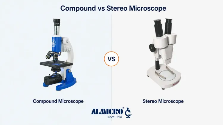
The main difference between a compound microscope and a stereo microscope comes down to the specimen you're examining. A compound microscope delivers high magnification for thin transparent samples such as cells and tissue sections. A stereo microscope gives a three-dimensional view of larger solid objects such as insects or circuit boards at comparatively lower magnification. Both instruments are optical microscopes but they serve different purposes. The compound microscope vs stereo microscope comparison comes down to magnification range, optical path, illumination method and the type of specimen each handles best. Choosing the right one depends on what you need to observe and how you plan to work with the sample.
Compound Microscope vs Stereo Microscope: Quick Comparison
| Feature | Compound Microscope | Stereo Microscope |
|---|---|---|
| Typical magnification | Approximately 40x–1000x | Commonly 7x–45x, depending on the model |
| Image appearance | Primarily two-dimensional | Three-dimensional |
| Specimen type | Thin and transparent | Solid, larger and opaque |
| Illumination | Mainly transmitted light | Mainly reflected light |
| Optical path | One primary image path | Two separate optical paths |
| Working distance | Short | Long |
| Sample preparation | Usually required | Usually minimal |
| Depth of field | Lower | Greater |
| Common uses | Cells, tissues and microorganisms | Insects, circuit boards and gems |
| Object manipulation | Limited | Possible during observation |
Specifications vary between models and configurations, so always check the exact spec sheet for a particular instrument.
What Is a Compound Microscope?
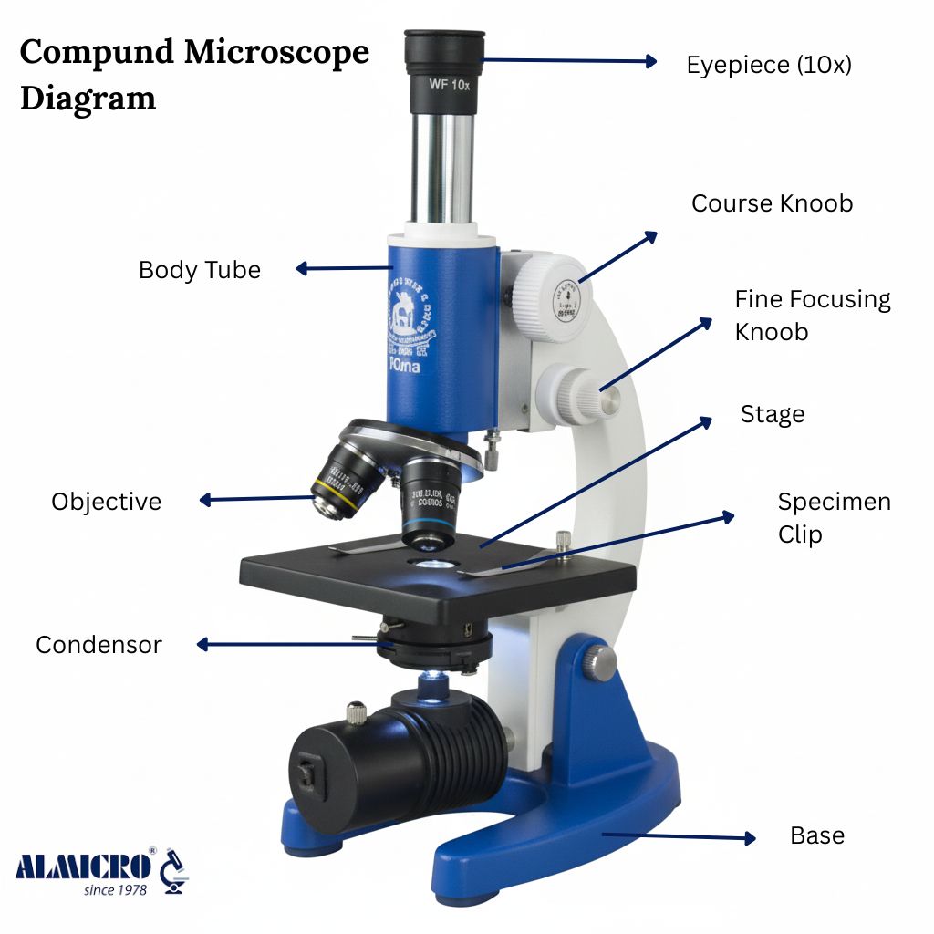
A compound microscope uses two lens systems working together, an objective lens near the specimen and an eyepiece near your eye. These lenses combine their magnifying power to produce images far beyond what a single lens could achieve. Because of this design, a compound microscope is mainly used for specimens mounted on glass slides rather than solid objects you can hold. Light usually passes upward through the specimen from a lamp positioned below the stage, which is why this method is called transmitted illumination. The image that reaches your eye is generally flat and two-dimensional. This makes the compound microscope the standard choice wherever fine internal detail matters more than surface texture, from a school biology lab to a hospital pathology department.
Compound Microscope Magnification
Total magnification is calculated by multiplying the eyepiece magnification by the objective magnification.
Total magnification = Eyepiece magnification × Objective magnification
Example: a 10x eyepiece paired with a 40x objective gives 400x total magnification.
Most compound microscopes include a set of standard objectives:
- 4x
- 10x
- 40x
- 100x oil immersion
Switching between these objectives lets a single instrument cover a wide compound microscope magnification range for different specimen types.
Common Uses of a Compound Microscope
- Examining cells
- Blood smear observation
- Tissue section examination
- Studying microorganisms
- Biology practicals
- Pathology and microbiology laboratories
These compound microscopes span classrooms, diagnostic labs and research facilities where cellular detail is essential.
Compound Microscope Diagram :
What Is a Stereo Microscope?
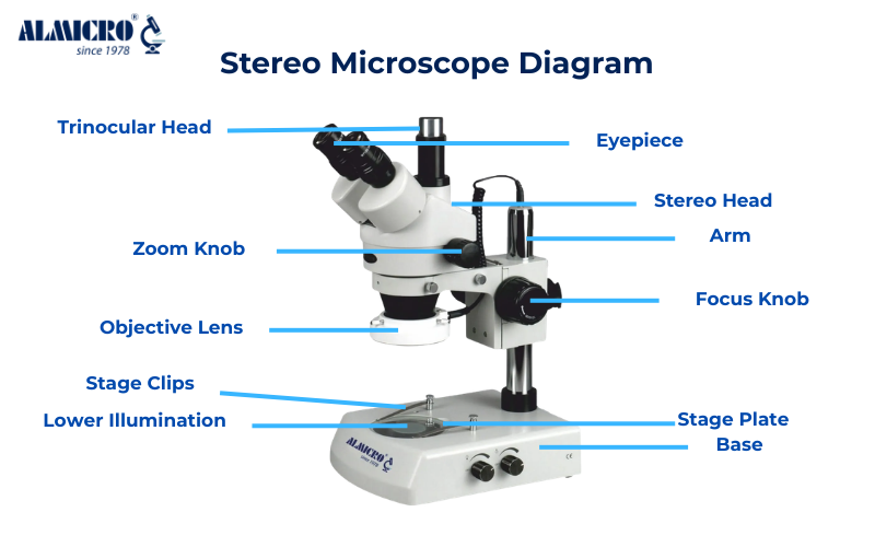
A stereo microscope, also called a stereoscope or dissecting microscope, uses two separate optical paths instead of one. Each eye receives a slightly different angle of the same object, and your brain combines these two views into a three-dimensional image. This is what gives a stereo microscope its depth perception, something a standard compound microscope cannot reproduce. Magnification is lower than a compound microscope but the working distance is much longer, leaving room to move tools, tweezers or your hands beneath the lens. Most specimens can be placed directly under a stereo microscope without any slide preparation. This combination of depth, working space and minimal setup is why the stereo microscope has become a fixture in dissection, electronics and quality control work.
Stereo Microscope Magnification
Stereo microscopes generally operate at lower magnification than compound microscopes. The exact stereo microscope magnification depends on the eyepiece, objective and zoom system fitted to the instrument. Two configurations are common:
- Fixed-power stereo microscopes with a set magnification
- Zoom stereo microscopes that allow continuous adjustment within a range
Common Uses of a Stereo Microscope
- Biological dissection
- Insect examination
- Electronics and PCB inspection
- Jewellery and gemstone observation
- Watch and small component repair
- Surface inspection
- Industrial quality control
These stereo microscopes all share one requirement, a clear three-dimensional view of a solid object rather than a thin slide.
Stereo Microscope Diagram:
10 Key Differences Between Compound and Stereo Microscopes
Here is a closer look at the difference between compound microscope and stereo microscope across the factors that matter most when choosing an instrument.
1. Magnification
Compound microscopes provide much higher magnification, often reaching several hundred times or more. Stereo microscopes provide lower magnification but cover a wider viewing area, which suits larger objects better.
2. Image Appearance
A compound microscope produces a primarily two-dimensional image. A stereo microscope produces a three-dimensional view with real depth perception.
3. Optical Path
A compound microscope directs one primary image toward the eyepieces. A stereo microscope uses two separate optical paths that create slightly different viewing angles for each eye.
4. Specimen Type
Compound microscopes work best with cells, tissues, microorganisms and prepared slides. Stereo microscopes suit insects, rocks, circuit boards, gems and mechanical parts.
5. Illumination
Compound microscopes mainly rely on transmitted light passing up through the specimen. Stereo microscopes mainly use reflected light falling on the specimen from above, though some stereo models also support transmitted illumination for translucent samples.
6. Working Distance
Working distance is the space between the objective lens and the specimen. A compound microscope has a shorter working distance while a stereo microscope has a longer one. That extra space under a stereo microscope allows tools and hands to work beneath the objective while observing.
7. Sample Preparation
Compound microscope specimens usually need to be mounted, stained or sliced thin enough for light to pass through. Stereo microscope specimens can usually be placed under the lens and observed directly.
8. Field of View
Compound microscopes show a smaller area at high magnification. Stereo microscopes provide a wider field of view, useful when scanning an entire object rather than a single detail.
9. Depth of Field
Compound microscopes have a comparatively shallow depth of field so only a thin plane stays in sharp focus. Stereo microscopes keep more of an uneven specimen surface in focus at once.
10. Applications
Compound microscopes are primarily used in education, biology, pathology and research settings. Stereo microscopes are used in dissection, electronics, manufacturing and surface inspection work.
Difference Between Compound Microscope and Dissecting Microscope
A dissecting microscope is simply another name commonly used for a stereo microscope. It offers a low-magnification three-dimensional view along with a working distance long enough to dissect or manipulate a specimen while watching it under the lens. This makes the compound microscope vs dissecting microscope comparison essentially the same as compound versus stereo. A compound microscope, on the other hand, provides much higher magnification for thin specimens mounted on glass slides. It comes into play when cellular or microscopic detail is required rather than a surface-level view of a whole object.
Binocular Compound Microscope vs Stereo Microscope
This is a common point of confusion. A binocular compound microscope has two eyepieces for more comfortable viewing, but both eyepieces generally receive the same primary image from a single optical path. A stereo microscope, by contrast, uses two genuinely separate optical paths, one for each eye. Simply having two eyepieces does not make an instrument stereoscopic. A binocular compound microscope still produces a primarily flat 2D image, while a true stereo microscope delivers real depth perception through its dual optical design.
What Can You See With Each Microscope?
| Specimen or Task | Recommended Microscope |
|---|---|
| Blood cells | Compound Microscope |
| Prepared tissue section | Compound Microscope |
| Microorganisms | Compound Microscope |
| Onion epidermal cells | Compound Microscope |
| Whole insect | Stereo Microscope |
| Plant dissection | Stereo Microscope |
| Circuit board | Stereo Microscope |
| Gemstone surface | Stereo Microscope |
| Small mechanical part | Stereo Microscope |
Which Microscope Should You Choose?
Choose a compound microscope for observing cells, tissues, microorganisms and prepared slides. Choose a stereo microscope for solid objects, surface inspection, dissection and any task that needs depth perception.
Choose a Compound Microscope When:
- You need higher magnification
- Your specimen is thin or transparent
- You need to study cellular structures
- Your specimens are mounted on slides
- Your work involves biology, pathology or microbiology
Choose a Stereo Microscope When:
- You need a three-dimensional surface view
- Your object is solid or opaque
- You need a longer working distance
- You must handle the object while observing it
- Your work involves dissection, electronics, gems or inspection
When Might You Need Both?
- Biology laboratories conducting slide examination and dissection
- Educational institutions covering multiple practical activities
- Research facilities studying both internal structures and external surfaces
Choosing a Microscope for Indian Schools, Laboratories and Industries
A few factors decide which instrument fits your setup best:
- Specimen type
- Required magnification
- Optical quality
- Working distance
- Fixed or zoom configuration
- Monocular, binocular or trinocular head
- LED illumination
- Mechanical stage requirements
- Camera compatibility
- Spare part availability
- Warranty and technical support
ALMICRO is the microscope and laboratory equipment brand of Micro Measure & Instruments, manufactured in Ambala Cantt, Haryana since 1978. Its compound, stereo and research microscopes are supplied to schools, hospitals, research institutes and industrial units across India, built to match the specimen types and working conditions described above.
Final Summary
A compound microscope offers higher magnification, works with prepared slides and reveals microscopic detail invisible to the naked eye. A stereo microscope offers lower magnification, a genuine 3D view and works well with solid specimens that need no preparation. Neither instrument is universally better, they're built for different tasks. The right choice always comes back to your specimen and what you need to observe. For labs and institutions that need both capabilities, ALMICRO manufactures compound and stereo microscopes suited to Indian classrooms, hospitals and industrial settings.
Learn More About Microscopes
Explore these related ALMICRO microscope guides:
- Compound Microscope: Parts, Diagram, Features & Uses
- Stereo Microscope: Principle, Diagram, Uses & Parts
- Types of Microscopes: Working, Uses and Diagrams
- Microscope Objective Lens: Types and Magnification Guide
- Monocular vs Binocular Microscope: Which Should You Choose?
Shop Our Best Sellers in Compound Microscopes and Stereo Microscopes:
Explore Our best-selling compound microscope and stereo microscopes designed for educational, medical, research and industrial applications.